Huid-Darm As: Wat hebben de darmen met onze huid te maken?

De huidepidermis, samen met zijn aanhangsels zoals zweet- en talgklieren, biedt een totaal huidoppervlak van ongeveer 25 m2 en is een van de grootste epitheeloppervlakken voor interactie met microben [1]. De huid is een barrière van de eerste lijn vanuit de externe omgeving en staat er continu mee in contact. Het gastro-intestinale (GI) kanaal is een van de grootste interfaces (30 m2) tussen de gastheer en zijn omgeving [2]. Er wordt geschat dat ongeveer 60 ton voedsel in een mensenleven door het darmkanaal gaat, wat allemaal een grote invloed heeft op de menselijke gezondheid [3]. Zowel de darm als de huid zijn sterk doordrongen van microbiota, waarbij wordt geschat dat de huid ongeveer 1012 microbiele cellen heeft, terwijl de darm goed is voor 1014 microbiele cellen [4,5]. De microbiota wijzen op de verzameling van specifieke micro-organismen die aanwezig zijn binnen een bepaalde omgeving. De opkomst van next-generation sequencing in het afgelopen decennium heeft ongekende inzichten geboden in de samenstelling van het microbioom, zowel op de huid als in de darm. Het microbioom verwijst naar de genomen aanwezig in een bepaalde omgeving, wat betekent dat het de ophoping is van al hun genetisch materiaal (d.w.z. DNA en RNA). Beide organen worden gekenmerkt door een lage microbiële diversiteit op fylum-niveau, maar een hoge diversiteit op soortniveau [6]. Het microbioom biedt de gastheer tal van voordelen, zoals het vormen van het immuunsysteem, bescherming tegen pathogenen, afbraak van metabolieten en het behoud van een gezonde barrière [3].

De immuun-modulerende potentie van het microbioom op verre orgaanlocaties is een groeiend onderzoeksgebied. Vooral de invloed van het darmmicrobioom op verre organen, zoals de longen, hersenen en huid, heeft de volgende onderzoeksgebieden gecreëerd: darm-long as, darm-hersen as en darm-huid as [7]. Het aangeboren en adaptieve immuunsysteem verandert de microbiële samenstelling; echter, het lokale microbioom kan ook het immuunsysteem moduleren. De onderliggende mechanismen van hoe het darmmicrobioom het immuunsysteem van de huid verandert, en vice versa, worden momenteel onderzocht. Verschillende huidpathologieën worden gepresenteerd als darmcomorbiditeiten. Verschillende studies hebben de tweerichtingsverbinding tussen darmdysbiose en onevenwichtigheden in de huidhomeostase aangetoond, met een specifieke rol van darmmicrobiota-dysbiose in de pathofysiologie van meerdere inflammatoire ziekten [8,9,10].

Een samenvatting van recente bevindingen in het huid- en darmmicrobioom bij meerdere huidaandoeningen staat in deze review, waarbij enkele potentiële mechanismen worden belicht die ten grondslag liggen aan de darm-huid as.


Huid versus Darm Barrière

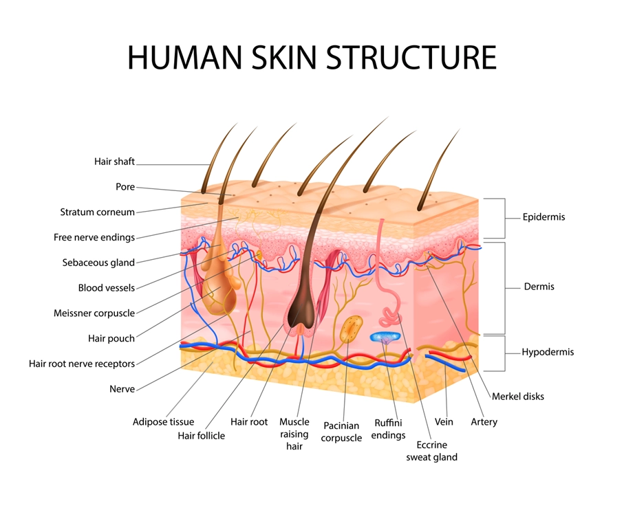
De darm- en huidbarrière delen opvallend veel kenmerken. De darm en huid zijn sterk analoog aan elkaar qua doel en functionaliteit. Beide organen zijn sterk geïnnerveerd en gevaccineerd, omdat ze beiden essentieel zijn voor immuun- en neuro-endocriene functies [11]. De darm-huid as ontstaat door deze gelijkenis [11]. Het binnenoppervlak van de darm en het buitenoppervlak van de huid worden beide bedekt door epitheelcellen (EC’s) die rechtstreeks contact hebben met de exogene omgeving [12]. Op deze manier wordt het immuunsysteem continu geprikkeld om schadelijke en gunstige stoffen te onderscheiden. De activatie van immuuncellen begint al vroeg in het leven en vormt de basis voor tolerantie, een cruciaal concept waarvan wordt verondersteld dat het defect is bij verschillende auto-immuunziekten [13]. EC’s onderhouden een belangrijke schakel tussen het interne lichaam en de externe omgeving. Ze fungeren als een eerste verdedigingslinie om de binnenkomst van micro-organismen te voorkomen [12]. Keratine, dat aanwezig is in het gestratificeerde squameuze epitheel van de huid, vormt een formidabele fysieke barrière voor de meeste micro-organismen [14]. Bovendien maakt dit bestanddeel de huid bestand tegen zwakke zuren en basen, bacteriële enzymen en toxines [15]. Slijmvliezen bieden vergelijkbare mechanische barrières, omdat het bestaat uit een glycoproteïnelaag bovenop het epitheel waar commensale bacteriën verblijven [16,17]. De epitheelmembranen produceren beschermende chemicaliën die micro-organismen elimineren [18]. De zuurgraad van de huid (pH van 5,4 tot 5,9) creëert een onherbergzame omgeving voor potentiële pathogenen en remt bacteriële groei [19]. Talg, geproduceerd door de talgklieren, fungeert als een afdichting voor haarfollikels en bevat verschillende antimicrobiële moleculen en specifieke voedingslipiden voor gunstige micro-organismen [20,21]. Ondertussen bevatten speeksel en traanvocht lysozymen, gevolgd door de maagslijmvliezen die sterke zuren en eiwitverterende enzymen afscheiden [22]. Bovendien vangt slijm micro-organismen die het spijsverterings- en ademhalingssysteem binnendringen [23].

De tweede verdedigingslinie bestaat uit antimicrobiële peptiden (AMP’s), fagocyten en aangeboren lymfoïde cellen (ILC’s) [24]. Deze twee eerste verdedigingslinies vormen het aangeboren immuunsysteem [23]. AMP’s geproduceerd door keratinocyten, zoals cathelicidin en psorasin, bieden een effectieve barrièrefunctie voor de huid [25,26]. De serineprotease Kallikrein 5 (KLK5) splijt cathelicidin in actieve peptiden, zoals LL-37 [27]. Vergeleken met de huid varieert de samenstelling van de intestinale epitheelbarrière gedurende het maagdarmkanaal. Het proximale deel van het maagdarmkanaal, de mond en de slokdarm, is analoog aan de huid, bedekt met meerdere lagen van squameus epitheel, dat wordt gereinigd door slijm van speekselklieren en andere klieren [28]. Het overgebleven deel van het spijsverteringskanaal omvat een enkele laag actieve cellen, bijv. slijmbekercellen (slijmsecretie), enterochromaffinecellen (hormoonsecretie), enterocyten of colonocyten (absorptie), etc. [29,30]. Het intestinale epitheel bestaat uit een enkele laag enterocyten of colonocyten, en de integriteit van de barrière wordt beschermd door het immuunsysteem. De absorberende functionaliteit van de enterocyten in de dunne darm leidt tot een onderbroken laag slijm met minder slijmbekercellen [31]. Paneth-cellen zijn verrijkt in de crypten van de dunne darm die AMP’s afscheiden, die in de complexe slijmlaag worden geïntegreerd [32].

Microbiële geassocieerde moleculaire patronen (MAMP’s) worden bemonsterd via antigeenopname door membranen (M) cellen en slijmbekercellen naar dendritische cellen (DC’s), samen met directe transepitheliale lumen DC’s. Microbiële signalen worden waargenomen door ROR𝛾t aangeboren lymfoïde C-cellen (groep 3 ILC’s) die interleukine-17 (IL-17) en IL-22 produceren [33]. De laatste werkt rechtstreeks op de intestinale epitheelcellen (IEC’s) en activeert schadeherstelmechanismen, AMP’s en mucinegenen [34]. Plasmacellen in Peyer’s patches, gestimuleerd door DC’s, produceren IgA in het lamina propria op een T-cel-onafhankelijke manier [35,36]. De dikke darm daarentegen bevat een dikke, continue slijmlaag om de microbiota te compartimenteren, waarbij IgA en AMP’s een secundaire rol spelen [17]. De controle van immunologische processen binnen slijmvliezen is afhankelijk van de interactie tussen EC’s en DC’s, aangezien beide celtypen betrokken zijn bij het waarnemen en bemonsteren van antigenen [12]. Zowel in de huid als in de darm worden pathogenen bemonsterd via mechanismen die niet afhankelijk zijn van M-cellen [37]. De enige DC’s die binnen de epidermis worden gevonden, zijn de Langerhans-cellen (LC’s) [12].

Andere overeenkomsten tussen de darm en huidweefsels zijn de hoge celvernieuwingsgraad, wat hechting en infectie door de koloniserende microbiota remt [38,39]. De huid en de darm zijn de twee belangrijkste niches die prokaryotische en eukaryotische symbiotische micro-organismen herbergen [40,41]. Echter, de residente microbiota zijn vaak betrokken en spelen een cruciale rol bij de pathogenese van verschillende ziekten [42]. Beide weefsels reageren sterk op stress en angst, omdat ze vergelijkbare uitdagingen tegenkomen.

Opmerkelijk genoeg omvatten ziekten zoals inflammatoire darmziekte (IBD) en psoriasis een disfunctie van de epitheelbarrière en een verhoogde omzettingsgraad van epitheelcellen. De verhoogde permeabiliteit van de epidermale huid en de intestinale barrière komt door de versterkte interactie van allergenen en pathogenen met ontstekingsreceptoren van immuuncellen. Beide ziekten hebben een vergelijkbare immuunrespons en betrekken fagocyten, dendritische en natuurlijke killercellen, samen met een reeks cytokinen en AMP’s die een T-celrespons induceren [43]. Bovendien worden beide ziekten gekenmerkt door dysbiose in de samenstelling van het microbiële ecosysteem dat de respectieve interfaceranden bedekt [43].

Het darmmicrobioom is het grootste endocriene orgaan en produceert minstens 30 hormoonachtige verbindingen, zoals korteketenvetzuren (SCFA’s), secundaire galzuren, cortisol en neurotransmitters zoals gamma-aminoboterzuur (GABA), serotonine, dopamine en tryptofaan. Bepaalde leden van het darmmicrobioom reageren op hormonen die door de gastheer worden uitgescheiden [44]. De hormoonachtige pleiotropische verbindingen die door het darmmicrobioom worden geproduceerd, komen in de bloedbaan terecht en kunnen op afgelegen organen en systemen werken, zoals de huid [44]. Talrijke studies hebben bewijs geleverd voor een diepgaande tweezijdige relatie tussen gastro-intestinale gezondheid en huidhomeostase door aanpassing van het immuunsysteem [45,46,47]. Modulatie van het immuunsysteem vindt voornamelijk plaats via het darmmicrobioom. Echter, commensale huidmicrobiota zijn even essentieel voor het behoud van de immuunhomeostase van de huid [48]. Zowel de darm als de huid herbergen diverse bacteriële, schimmel- en virale soorten die symbiose met de menselijke omgeving behouden. Het verstoren van deze balans kan leiden tot een verminderde barrièrefunctie. Het herstel van de huidhomeostase na verstoring of stress via het darmmicrobioom heeft invloed op zowel aangeboren als adaptieve immuniteit.


Betrokkenheid van de huid- en darmmicrobiomen

De huid is de grootste en meest externe barrière van het lichaam met de buitenomgeving. Het is rijk aan immuuncellen en zwaar gekoloniseerd door microbieel leven, dat op zijn beurt de immuuncellen traint en het welzijn van de gastheer bepaalt [49]. Het huidmicrobioom heeft de afgelopen jaren aanzienlijke aandacht gekregen in de dermatologie, huidaandoeningen en de verbinding en invloed op het immuunsysteem. Veel huidaandoeningen worden geassocieerd met een onevenwicht in het huidmicrobioom (Tabel 1). Steeds meer studies tonen verrijkte pathogenen en microbiota die geassocieerd worden met huidaandoeningen, sommige zijn voor de hand liggend en andere verrassender. Het is echter moeilijk vast te stellen of het veranderde huidmicrobioom een oorzaak of gevolg is van de huidaandoening.”

Hieronder bieden we een overzicht van negen veelvoorkomende huidaandoeningen en hun respectievelijke pathofysiologie, en de kennis over de verstoringen in hun huidmicrobioom, een verstoord darmmicrobioom, en/of de relatie met een specifiek dieet.


Acne Vulgaris
Acne Vulgaris (Acne): Acne Vulgaris is een complexe aandoening waarbij verschillende factoren samenkomen om het te veroorzaken. De balans van de huidmicrobiota, hormonale schommelingen, talgproductie, dieetinvloeden en immuunpaden spelen allen een belangrijke rol bij het ontstaan van deze aandoening.

De huidmicrobiota speelt een cruciale rol in de pathologie van Acne Vulgaris, vooral bepaalde stammen van Cutibacterium acnes. De verstoring in de huidmicrobiota is geassocieerd met diverse factoren, zoals de inductie van talg, directe stimulatie van het immuunsysteem, diversiteit binnen de C. acnes populatie, porfyrineproductie, mobiele genetische elementen, CRISPR/CAS loci en de productie van korteketenvetzuren (SCFAs). Ondanks uitgebreid onderzoek blijft de precieze choreografie van deze pathogene gebeurtenissen enigszins ongrijpbaar.

Interessant genoeg laten studies duidelijke verschillen zien in de huidmicrobiota van acne patiënten, met een lagere aanwezigheid van gunstige bacteriën en een toename van bepaalde bacteriestammen die met de aandoening geassocieerd worden. Deze disbalans beperkt zich niet tot de huid; onderzoek wijst op veranderingen in de diversiteit van de darmmicrobiota bij acne patiënten, wat duidt op een mogelijk verband tussen darmgezondheid en de ontwikkeling of ernst van acne.

De relatie tussen acne en dieet benadrukt verder de complexiteit van deze aandoening. Diëten met een hoge glycemische lading, verzadigde vetten en specifieke vetzuurinname worden geassocieerd met de ernst van acne. Deze voedingscomponenten verstoren mogelijk signaalpaden voor voedingsstoffen, wat leidt tot ongecontroleerde stimulatie van bepaalde biologische processen zoals de synthese van vetzuren en triglyceriden in talg. Deze veranderingen kunnen een omgeving bevorderen die gunstig is voor de groei van Cutibacterium acnes, waardoor acne-symptomen verergeren.

Therapeutische benaderingen omvatten voornamelijk antibacteriële middelen, retinoïden of comedolytische agentia. De multifactoriële aard van acne vereist echter een begrip dat de onderliggende ziekteprocessen meerdere bijdragende factoren omvatten. Gepersonaliseerde behandelingen die rekening houden met de complexe interactie tussen huidmicrobiota, hormonale balans, dieetinvloeden en immuunreacties bieden hoop op effectieve behandeling van deze aandoening.

Atopische Dermatitis (AD): Atopische Dermatitis, een ontstekingsziekte van de huid, is het resultaat van een complex samenspel tussen genetische aanleg, omgevingsfactoren en immuunreacties. De pathofysiologie omvat een verstoorde huidbarrière, chronische ontsteking en microbiële dysbiose, vooral in de huidmicrobiota.

De microbiële dysbiose van de huid bij AD vertoont veranderingen gekenmerkt door een verminderde bacteriële diversiteit en een verhoogde aanwezigheid van Staphylococcus aureus, terwijl gunstige bacteriën zoals Cutibacterium, Corynebacterium, Streptococcus, Acinetobacter, Prevotella en Malassezia afnemen. Deze disbalans heeft een significante invloed op de inflammatoire reacties die bij AD optreden, vooral door de groei en pathogenese van Staphylococcus aureus.

Bovendien is darmdysbiose bij AD-patiënten een onderwerp van aanzienlijke interesse. Studies suggereren dat patiënten met AD een verminderde diversiteit van de darmmicrobiota vertonen, met specifieke variaties in de abundantie van verschillende bacteriesoorten in vergelijking met gezonde controles. Probiotische interventies hebben belofte getoond in het beheer van AD, wat wijst op het potentieel om de darmmicrobiota te beïnvloeden om immuunreacties bij huidaandoeningen te beïnvloeden.

De correlatie tussen dieet en AD benadrukt verder de veelzijdige aard van deze aandoening. Verminderde consumptie van fruit, groenten en omega-3 vetzuren, samen met een verhoogde inname van omega-6 vetzuren, zijn geassocieerd met AD. Deze voedingsgewoonten kunnen ontstekingen bevorderen en de ernst van AD beïnvloeden, hoewel verder onderzoek vereist is om het definitieve effect van dieetmanipulaties op deze aandoening te bevestigen.

Psoriasis: Psoriasis, een immuun-gemedieerde inflammatoire aandoening, kenmerkt zich door roodheid, schilfering en verdikte huidletsels op verschillende delen van het lichaam. Deze aandoening wordt beïnvloed door genetische factoren, levensstijl en omgeving, en wordt beschouwd als een complexe ziekte met systemische gevolgen.

Het immuungemedieerde karakter van psoriasis draait voornamelijk om de Th17-route, waarbij ontstekingen worden veroorzaakt door IL-23/IL-17-gemedieerde reacties, terwijl TNF het ontstekingsproces versterkt. Hoewel er vooruitgang is geboekt in het begrijpen van de immunologische basis, blijft de exacte oorzaak van psoriasis onduidelijk. De aanwezigheid van een antivirale respons in psoriasis, gesuggereerd door de aanwezigheid van type I interferonen, wijst op mogelijke betrokkenheid van de huidmicrobiota bij virale reacties.

Studie naar de psoriatische huidmicrobiota onthult een gevarieerd beeld, met verschillende uitkomsten met betrekking tot microbiële diversiteit en soortenrijkdom. De huid van psoriasispatiënten vertoont vaak een veranderde bacteriële samenstelling, met een hogere aanwezigheid van Staphylococcus- en Streptococcus-soorten en een verminderde diversiteit in bepaalde microbiële groepen. Daarnaast is darmdysbiose in verband gebracht met psoriasis, wat mogelijk de ernst van de aandoening beïnvloedt en de respons op behandelingen kan beïnvloeden.

De complexe relatie tussen psoriasis en darmgezondheid wordt duidelijk door de associaties met intestinale immuunstoornissen, zoals IBD, UC en coeliakie. Structurele afwijkingen in de darm en veranderingen in de darmintegriteit zijn gerapporteerd bij psoriasispatiënten, wat wijst op systemische implicaties die verder gaan dan alleen de huidverschijnselen. Behandelingen voor psoriasis, inclusief systemische therapieën, hebben invloed op de darmmicrobiota, wat wijst op potentiële interventiemogelijkheden.

Levensstijl en dieetfactoren hebben aanzienlijke invloed op psoriasis. Roken, alcoholgebruik en obesitas zijn gekoppeld aan verergering van huidletsels en respons op behandelingen. Aan de andere kant hebben gezonde eetgewoonten, inclusief gewichtsmanagement en specifieke dieetregimes zoals een caloriearm ketogeen dieet of intermitterend vasten, potentiële voordelen laten zien bij het beheren van de ernst van psoriasis.

De samenvattingen van deze aandoeningen onderstrepen de complexe wisselwerking tussen huidaandoeningen, microbiële veranderingen, immuunreacties en dieetinvloeden. Een dieper begrip van deze veelzijdige verbanden is cruciaal voor de ontwikkeling van gerichte interventies en gepersonaliseerde therapieën om deze chronische aandoeningen effectief te beheren.

Hidradenitis Suppurativa (HS): Deze chronische huidaandoening, die ongeveer 0,3% van de wereldbevolking treft, manifesteert zich op plaatsen waar de huid tegen elkaar wrijft of waar zweetklieren aanwezig zijn, zoals de oksels, liezen of onder de borsten. HS veroorzaakt pijnlijke, terugkerende bulten die kunnen openspringen en tunnels onder de huid kunnen vormen. Hoewel de exacte oorzaak onduidelijk blijft, zijn er sterke aanwijzingen voor een verband met het immuunsysteem en ontstekingsreacties. Onderzoek suggereert dat genetica, hormonale factoren en levensstijlgewoonten zoals roken en obesitas mogelijk bijdragen aan de ontwikkeling ervan.

Rosacea: Rosacea is een chronische huidaandoening gekenmerkt door roodheid, zichtbare bloedvaten en soms kleine rode bultjes op het gezicht. Hoewel de exacte oorzaak niet volledig wordt begrepen, wordt aangenomen dat het een combinatie is van genetische en omgevingsfactoren. Bepaalde triggers zoals blootstelling aan de zon, pikant voedsel, alcohol, stress en sommige huidverzorgingsproducten kunnen de symptomen verergeren. Cathelicidine, een eiwit betrokken bij de immuunafweer van het lichaam, lijkt overmatig tot expressie te komen in rosacea-gevoelige huid, wat leidt tot ontsteking en verwijding van bloedvaten. De huidmicrobiota, vooral een overgroei van bepaalde mijten en bacteriën, kan ook bijdragen aan de ontwikkeling en verergering ervan.

Roos en Seborroïsch Eczeem: Roos, een veelvoorkomende hoofdhuidaandoening gekenmerkt door schilfering en jeuk, wordt vaak geassocieerd met de aanwezigheid van een schimmel genaamd Malassezia. Deze gistachtige schimmel is een normaal onderdeel van de hoofdhuidmicrobiota, maar een overgroei ervan kan leiden tot irritatie en schilfering. Seborroïsch eczeem is een ernstigere vorm van roos die gepaard gaat met rode, ontstoken huid en zich kan uitbreiden buiten de hoofdhuid naar andere gebieden met talgklieren, zoals het gezicht en bovenlichaam. Beide aandoeningen kunnen verband houden met onevenwichtigheden in de microbiële gemeenschap van de huid, vooral een toename van bepaalde schimmel- of bacteriesoorten. Dieet en gezondheid van de darmen lijken ook een rol te spelen, waarbij enig bewijs een verband legt tussen suikerinname en darmgerelateerde problemen en de ernst van de symptomen.

Alopecia: Alopecia areata veroorzaakt plotseling haarverlies in kleine, ronde plekken op de hoofdhuid of andere delen van het lichaam. Het wordt beschouwd als een auto-immuunziekte waarbij het immuunsysteem van het lichaam per ongeluk de haarzakjes aanvalt, waardoor ze krimpen en stoppen met haargroei. Hoewel genetica en disfunctie van het immuunsysteem als belangrijke factoren worden beschouwd, blijven de exacte triggers voor deze immuunrespons onduidelijk. Onderzoek onderzoekt de rol van de huidmicrobiota bij deze aandoening, waarbij wordt vermoed dat een onevenwicht in bepaalde micro-organismen op de hoofdhuid zou kunnen bijdragen aan het ontstaan of de progressie ervan. Bovendien suggereert enig bewijs dat de gezondheid van de darmen en de samenstelling van de darmmicrobiota van invloed kunnen zijn op de ontwikkeling of ernst van alopecia. Voedingsstoffentekorten, met name vitamines en mineralen die essentieel zijn voor een gezonde haargroei, spelen ook een rol, wat wijst op een potentieel verband tussen dieet en het beheer van alopecia.


Huidkanker

Huidkanker is een veelvoorkomende maligniteit die kan worden onderverdeeld in twee categorieën: invasieve melanoom, waarbij melanocyten ongecontroleerd delen, en niet-melanoom huidkankers (NMSC’s). De laatste omvat tumoren met een keratinocytaire oorsprong, zoals basaalcelcarcinoom (BCC) en plaveiselcelcarcinoom (SCC) [299]. Er zijn verschillende risicofactoren die kunnen leiden tot melanoom en NMSC’s, waaronder constitutionele aanleg, immuunsuppressieve status en blootstelling aan omgevingsrisicofactoren zoals ultraviolette straling [300]. Bovendien kunnen actinische keratose en Bowen’s ziekte ook leiden tot SCC [301]. De immunologie met betrekking tot cutane componenten werd de afgelopen decennia beter begrepen door mechanismen van immuunbewaking en het immunoediting raamwerk. De immunogeniteit van tumorcellen verandert door een gewijzigde expressie van (tumor-geassocieerde) antigenen, zoals verminderde MHC-1-expressie, wat resulteert in de ontwikkeling van maligniteit [302].

De impact van virussen en UV-straling op huidkanker is uitgebreid onderzocht. Onlangs werd een lagere incidentie van huidkanker ontdekt bij kiemvrije ratten. Hierdoor wordt vermoed dat een verstoorde huidmicrobiota kan leiden tot de ontwikkeling van verschillende vormen van huidkanker. Het is echter nog onduidelijk of tumorcellen of microbiële dysbiose de progressie veroorzaken [303]. Onderzoeken hebben de link onderzocht tussen verschillende vormen van huidkanker en dysbiose van het bacteriële huidmicrobioom bij ontstekingsziekten waarbij Th17 betrokken is, zoals psoriasis en acne [103]. Bovendien zijn SCC en actinische keratose onlangs ook geassocieerd met een toename van bepaalde stammen van S. aureus in combinatie met een afname van huidcommensalen [100]. Verder vertoonden melanoommonsters volgens een recent onderzoek door Mrázek et al. verhoogde niveaus van Fusobacterium- en Trueperella-genera [102]. Bovendien kan een toename van Merkelcel polyomavirus (MCPyV), een virus dat naar verluidt een aanhoudende bewoner van de huid is, leiden tot Merkelcelcarcinoom (MCC) [103]. Aan de andere kant bleek uit onderzoek dat specifieke S. epidermidis-stammen selectief de proliferatie van tumorcellijnen remmen en bescherming bieden tegen de voortgang van door UVB geïnduceerde huidpapillomen in preklinische modellen [304].

Patiënten met kanker worden vaak blootgesteld aan een verstoord darmmicrobioom als gevolg van therapieën die de samenstelling en immuniteit van deze microbiota beïnvloeden [306]. Hoewel de relatie tussen deze dysbiose en huidkanker specifiek nog onduidelijk is, is de relatie met kanker in het algemeen al enigszins onderzocht. Meer onderzoek is echter nodig om de correlatie tussen darmdysbiose en huidkanker te onderzoeken. Ten slotte is het onzeker of tumorontwikkeling secundair is aan bacteriële dysbiose.


Wondgenezing

Wondgenezing van de huid is een zeer complex en georganiseerd proces en bestaat uit overlappende fasen van acute genezing [307]. Meerdere celtypen, voornamelijk epidermale keratinocyten, neutrofielen en macrofagen, zijn betrokken en interageren met de aanwezige commensale microbiota. Wanneer een van deze fasen wordt belemmerd, zal de epitheliale barrière niet goed genezen en wordt een wond chronisch. Verstoorde wondgenezing is een grote uitdaging voor het gezondheidszorgsysteem en treft ongeveer 1% tot 2% van de bevolking in ontwikkelde landen [308,309]. De prevalentie van chronische wonden is hoger bij oudere mensen met onderliggende pathologieën zoals diabetes mellitus, vaatziekten en obesitas [310].

Wonden bieden een ideale omgeving voor microbiota om toegang te krijgen tot onderliggend weefsel en om te koloniseren en te groeien [318,319]. Commensale microbiota zijn naar verluidt gunstig voor het wondgenezingsproces. Ze zijn essentieel voor het reguleren van het huidinnate immuunsysteem en stimuleren de productie van antimicrobiële moleculen die bescherming bieden tegen intracellulaire pathogenen [318,320,321].

Veranderingen in het commensale huidmicrobioom kunnen bijdragen aan de vorming van chronische wonden. Recent onderzoek bij diermodellen suggereert dat probiotica niet-genezende wonden kunnen hinderen en genezen. Dit vormt een boeiend onderzoeksveld met veelbelovende toepassingen voor therapieën en cosmetica. Het begrijpen van de interacties tussen het microbioom en de gastheerweefsels zal leiden tot een beter begrip van gezondheid en ziekte en zal nieuwe mogelijkheden creëren. Er is behoefte aan goed ontworpen klinische onderzoeken om de effectiviteit van op microbiota gerichte behandelingen te beoordelen.


Conclusies

De huidaandoeningen zoals besproken in dit manuscript zijn het resultaat van een complexe interactie tussen genetische aanleg, levensstijl en het immuunsysteem. Meer specifiek staat dit laatste constant in verbinding met het zenuwstelsel en het endocriene systeem. Deze interacties stellen microbiota in staat een sleutelrol te spelen, vooral in organen zoals de huid en darmen die rijk zijn aan immunoregulatoren en microbiota. Bovendien suggereren observaties zoals de preventie van AD door probiotica en de verhoogde prevalentie van darmgerelateerde aandoeningen bij chronische huidaandoeningen dat huidaandoeningen kunnen worden gelinkt aan het maagdarmstelsel. De belangrijkste hypothese is gebaseerd op de gezondheid van de darm, die wordt gereguleerd door voedingsfactoren, gemedieerd via het darmmicrobioom en het immuunsysteem, wat leidt tot systemische effecten, inclusief huidgezondheid. De integriteit van de darmbarrière speelt hierbij een sleutelrol, maar het bestaan ervan blijft sterk bediscussieerd.

Deze dysbiose van het darmmicrobioom vormt een interessant onderzoeksgebied met toepassingsmogelijkheden. Pre- en probiotica gericht op het darmmicrobioom kunnen worden gebruikt om de huidgezondheid te beïnvloeden [45]. Muizen die probiotica kregen met Lactobacillus reuteri vertoonden bijvoorbeeld een glanzendere en dikkere vacht, gemedieerd door IL-10, en verbeterden ook het integumentaire systeem toen gezuiverde Foxp3+ T-cellen werden toegevoegd [329]. In een placebogecontroleerd onderzoek verkregen gezonde vrijwilligers een lagere transepidermale waterverlies en een lagere huidgevoeligheid bij consumptie van probiotica in vergelijking met de placebogroep [330]. Interessant genoeg hebben specifieke diëten, zoals caloriebeperking en vetarme diëten, ook verbeteringen in de darmepitheelbarrière of huidconditie laten zien, waaronder acne vulgaris, AD, psoriasis, wondgenezing, huidkanker en zelfs veroudering van de huid [339,340].

De aantrekkelijkheid van gerichte interventies op het darmmicrobioom via orale toediening lijkt omgekeerd evenredig te zijn met de complexiteit van het richten van de darm-huidas: het reguleren van het darmmicrobioom kan leiden tot systemische effecten, inclusief de huid en andere organen. Goed ontworpen klinische onderzoeken zijn nodig om de effectiviteit van op microbiota gerichte behandelingen te beoordelen. Recentelijk ontwikkelde sequencingtechnologieën zouden een gedetailleerd begrip van de mediators van het darm- en huidmicrobioom mogelijk moeten maken. Deze technologieën moeten worden ondersteund door biomarkeranalyses (zoals IgA, calprotectine en immuunmetingen) om de interactie tussen het microbioom en de integriteit van de darmbarrière te identificeren.

Hoewel het microbioom ook virale microbiota omvat, is er weinig bewijs beschikbaar over hoe virussen huidaandoeningen en de darmgezondheid beïnvloeden. Het aankomende volledige genoomonderzoek zou ons begrip van de rol van virussen in de darm-huidas moeten vergemakkelijken.

Huidgezondheidsresultaten moeten worden gedefinieerd om de impact te bepalen. Huidtapestrippen, dat recentelijk werd geïntroduceerd voor eiwit- en mRNA-kwantificatie, maakt niet-invasieve bemonstering mogelijk die minder belastend is voor de proefpersonen. Echter, huidtapestrippen onthult slechts beperkte informatie in vergelijking met huidbiopsieën.

Ten slotte moeten onderzoeksprotocollen deze complexiteit in overweging nemen: de kwaliteit van het vastleggen van voedingsgewoonten is cruciaal en is afhankelijk van de gekozen methode, zoals patiëntgerapporteerde uitkomstmaten (bijv. een voedselfrequentievragenlijst versus digitale apps voor calorieën tellen en voedingsdatabases). Bovendien moet het tijdstip van bemonstering zorgvuldig worden overwogen: proefpersonen kunnen bijvoorbeeld gevast zijn op het moment van bemonstering door het overslaan van ontbijt, in tegenstelling tot degenen die dat niet hebben gedaan. Bovendien moet rekening worden gehouden met de impact van het circadiane ritme in toekomstige klinische onderzoeken. De huidige literatuur ontbeert studies die de impact van het circadiane ritme van voedingsopname beschrijven. Het onderzoek van Parkar et al. beoordeelde het bewijs dat de effecten van veranderde slaap- en eetpatronen aantoont, die het gastheercircadiaansysteem kunnen verstoren en het darmmicrobioom kunnen beïnvloeden [341]. Naast ons eigen circadiane ritme, blijkt dat het microbioom ook een interne klok heeft die wordt gereguleerd door microbële metabolieten. Bepaalde voedingsstoffen worden vermoedelijk beter gemetaboliseerd op bepaalde momenten van de dag. Een hoge calorie-inname tijdens de avonduren wordt bijvoorbeeld geassocieerd met gewichtstoename, terwijl dezelfde calorie-inname tijdens de ochtenduren resulteert in gewichtsbehoud [345].

Kortom, de darm-huidas, met een centrale rol voor onze microbiota, vormt een boeiend onderzoeksgebied met veelbelovende therapeutische en cosmetische toepassingen. Het ontrafelen van de interacties tussen het microbioom en de gastheerweefsels zal leiden tot een beter begrip van gezondheid en ziekte en zal nieuwe mogelijkheden creëren. De noodzaak van goed ontworpen onderzoeken is van primair belang en vereist multidisciplinaire teams om samen te werken, in lijn met de samenwerking tussen ons eigen lichaam en microbiota.

Contact

Bron van de blog
  1. Gallo R.L. Human skin is the largest epithelial surface for interaction with microbes. J. Investig. Dermatol. 2017;137:1213–1214. doi: 10.1016/j.jid.2016.11.045. [PMC free article] [PubMed] [CrossRef] [Google Scholar]
  2. Helander H.F., Fändriks L. Surface area of the digestive tract–revisited. Scand. J. Gastroenterol. 2014;49:681–689. doi: 10.3109/00365521.2014.898326. [PubMed] [CrossRef] [Google Scholar]
  3. Thursby E., Juge N. Introduction to the human gut microbiota. Biochem. J. 2017;474:1823–1836. doi: 10.1042/BCJ20160510. [PMC free article] [PubMed] [CrossRef] [Google Scholar]
  4. Williams R. Benefit and mischief from commensal bacteria. J. Clin. Pathol. 1973;26:811. doi: 10.1136/jcp.26.11.811.[PMC free article] [PubMed] [CrossRef] [Google Scholar]
  5. Savage D.C. Microbial ecology of the gastrointestinal tract. Annu. Rev. Microbiol. 1977;31:107–133. doi: 10.1146/annurev.mi.31.100177.000543. [PubMed] [CrossRef] [Google Scholar]
  6. Grice E.A., Segre J.A. The skin microbiome. Nat. Rev. Microbiol. 2011;9:244–253. doi: 10.1038/nrmicro2537. [PMC free article] [PubMed] [CrossRef] [Google Scholar]
  7. Arck P., Handjiski B., Hagen E., Pincus M., Bruenahl C., Bienenstock J., Paus R. Is there a ‘gut–brain–skin axis’? Exp. Dermatol. 2010;19:401–405. doi: 10.1111/j.1600-0625.2009.01060.x. [PubMed] [CrossRef] [Google Scholar]
  8. Shah K.R., Boland C.R., Patel M., Thrash B., Menter A. Cutaneous manifestations of gastrointestinal disease: Part I. J. Am. Acad. Dermatol. 2013;68:189.e1–189.e21. doi: 10.1016/j.jaad.2012.10.037. [PubMed] [CrossRef] [Google Scholar]
  9. Thrash B., Patel M., Shah K.R., Boland C.R., Menter A. Cutaneous manifestations of gastrointestinal disease: Part II. J. Am. Acad. Dermatol. 2013;68:211.e1–211.e33. doi: 10.1016/j.jaad.2012.10.036. [PubMed] [CrossRef] [Google Scholar]
  10. Gloster H.M., Gebauer L.E., Mistur R.L.  Absolute Dermatology Review.Springer; Berlin/Heidelberg, Germany: 2016. Cutaneous manifestations of gastrointestinal disease; pp. 171–179. [Google Scholar]
  11. O’Neill C.A., Monteleone G., McLaughlin J.T., Paus R. The gut-skin axis in health and disease: A paradigm with therapeutic implications. BioEssays. 2016;38:1167–1176. doi: 10.1002/bies.201600008. [PubMed] [CrossRef] [Google Scholar]
  12. Shaykhiev R., Bals R. Interactions between epithelial cells and leukocytes in immunity and tissue homeostasis. J. Leukoc. Biol. 2007;82:1–15. doi: 10.1189/jlb.0207096. [PubMed] [CrossRef] [Google Scholar]
  13. Bach J.F. The effect of infections on susceptibility to autoimmune and allergic diseases. N. Engl. J. Med. 2002;347:911–920. doi: 10.1056/NEJMra020100. [PubMed] [CrossRef] [Google Scholar]
  14. Madison K.C. Barrier function of the skin:“la raison d’etre” of the epidermis. J. Investig. Dermatol. 2003;121:231–241. doi: 10.1046/j.1523-1747.2003.12359.x. [PubMed] [CrossRef] [Google Scholar]
  15. Lange L., Huang Y., Busk P.K. Microbial decomposition of keratin in nature—a new hypothesis of industrial relevance. Appl. Microbiol. Biotechnol. 2016;100:2083–2096. doi: 10.1007/s00253-015-7262-1. [PMC free article] [PubMed] [CrossRef] [Google Scholar]
  16. Pelaseyed T., Bergström J.H., Gustafsson J.K., Ermund A., Birchenough G.M., Schütte A., van der Post S., Svensson F., Rodríguez-Piñeiro A.M., Nyström E.E., et al. The mucus and mucins of the goblet cells and enterocytes provide the first defense line of the gastrointestinal tract and interact with the immune system. Immunol. Rev. 2014;260:8–20. doi: 10.1111/imr.12182. [PMC free article] [PubMed] [CrossRef] [Google Scholar]
  17. Kim Y.S., Ho S.B. Intestinal goblet cells and mucins in health and disease: Recent insights and progress. Curr. Gastroenterol. Rep. 2010;12:319–330. doi: 10.1007/s11894-010-0131-2. [PMC free article] [PubMed] [CrossRef] [Google Scholar]
  18. Janeway C.A., Jr., Travers P., Walport M., Shlomchik M.J.  Immunobiology: The Immune System in Health and Disease.5th ed. Garland Science; New York, NY, USA: 2001. The front line of host defense. [Google Scholar]
  19. Schmid-Wendtner M.H., Korting H.C. The pH of the skin surface and its impact on the barrier function. Skin Pharmacol. Physiol. 2006;19:296–302. doi: 10.1159/000094670. [PubMed] [CrossRef] [Google Scholar]
  20. Nakatsuji T., Kao M.C., Zhang L., Zouboulis C.C., Gallo R.L., Huang C.M. Sebum free fatty acids enhance the innate immune defense of human sebocytes by upregulating β-defensin-2 expression. J. Investig. Dermatol. 2010;130:985–994. doi: 10.1038/jid.2009.384. [PMC free article] [PubMed] [CrossRef] [Google Scholar]
  21. Dahlhoff M., Zouboulis C.C., Schneider M.R. Expression of dermcidin in sebocytes supports a role for sebum in the constitutive innate defense of human skin. J. Dermatol. Sci. 2016;81:124–126. doi: 10.1016/j.jdermsci.2015.11.013.[PubMed] [CrossRef] [Google Scholar]
  22. Patricia J.J., Dhamoon A.S. Physiology, Digestion. [(accessed on 2 November 2020)];2019 Available online: https://europepmc.org/books/nbk544242
  23. Brown E.M., Sadarangani M., Finlay B.B. The role of the immune system in governing host-microbe interactions in the intestine. Nat. Immunol. 2013;14:660–667. doi: 10.1038/ni.2611. [PubMed] [CrossRef] [Google Scholar]
  24. Spits H., Cupedo T. Innate lymphoid cells: Emerging insights in development, lineage relationships, and function. Annu. Rev. Immunol. 2012;30:647–675. doi: 10.1146/annurev-immunol-020711-075053. [PubMed] [CrossRef] [Google Scholar]
  25. Braff M.H., Zaiou M., Fierer J., Nizet V., Gallo R.L. Keratinocyte production of cathelicidin provides direct activity against bacterial skin pathogens. Infect. Immun. 2005;73:6771–6781. doi: 10.1128/IAI.73.10.6771-6781.2005. [PMC free article] [PubMed] [CrossRef] [Google Scholar]
  26. Gläser R., Harder J., Lange H., Bartels J., Christophers E., Schröder J.M. Antimicrobial psoriasin (S100A7) protects human skin from Escherichia coli infection. Nat. Immunol. 2005;6:57–64. doi: 10.1038/ni1142. [PubMed] [CrossRef] [Google Scholar]
  27. Yamasaki K., Schauber J., Coda A., Lin H., Dorschner R.A., Schechter N.M., Bonnart C., Descargues P., Hovnanian A., Gallo R.L. Kallikrein-mediated proteolysis regulates the antimicrobial effects of cathelicidins in skin. FASEB J. 2006;20:2068–2080. doi: 10.1096/fj.06-6075com. [PubMed] [CrossRef] [Google Scholar]
  28. Johansson M.E., Sjövall H., Hansson G.C. The gastrointestinal mucus system in health and disease. Nat. Rev. Gastroenterol. Hepatol. 2013;10:352. doi: 10.1038/nrgastro.2013.35. [PMC free article] [PubMed] [CrossRef] [Google Scholar]
  29. Cheng H., Leblond C. Origin, differentiation and renewal of the four main epithelial cell types in the mouse small intestine I. Columnar cell. Am. J. Anat. 1974;141:461–479. doi: 10.1002/aja.1001410403. [PubMed] [CrossRef] [Google Scholar]
  30. Podolsky D.K., Lynch-Devaney K., Stow J.L., Oates P., Murgue B., DeBeaumont M., Sands B.E., Mahida Y.R. Identification of human intestinal trefoil factor. Goblet cell-specific expression of a peptide targeted for apical secretion. J. Biol. Chem. 1993;268:6694–6702. doi: 10.1016/S0021-9258(18)53305-6. [PubMed] [CrossRef] [Google Scholar]
  31. Johansson M.E., Larsson J.M.H., Hansson G.C. The two mucus layers of colon are organized by the MUC2 mucin, whereas the outer layer is a legislator of host–microbial interactions. Proc. Natl. Acad. Sci. USA. 2011;108:4659–4665. doi: 10.1073/pnas.1006451107. [PMC free article] [PubMed] [CrossRef] [Google Scholar]
  32. Vaishnava S., Behrendt C.L., Ismail A.S., Eckmann L., Hooper L.V. Paneth cells directly sense gut commensals and maintain homeostasis at the intestinal host-microbial interface. Proc. Natl. Acad. Sci. USA. 2008;105:20858–20863. doi: 10.1073/pnas.0808723105. [PMC free article] [PubMed] [CrossRef] [Google Scholar]
  33. Qiu J., Heller J.J., Guo X., Zong-ming E.C., Fish K., Fu Y.X., Zhou L. The aryl hydrocarbon receptor regulates gut immunity through modulation of innate lymphoid cells. Immunity. 2012;36:92–104. doi: 10.1016/j.immuni.2011.11.011.[PMC free article] [PubMed] [CrossRef] [Google Scholar]
  34. Sonnenberg G.F., Monticelli L.A., Elloso M.M., Fouser L.A., Artis D. CD4+ lymphoid tissue-inducer cells promote innate immunity in the gut. Immunity. 2011;34:122–134. doi: 10.1016/j.immuni.2010.12.009. [PMC free article] [PubMed] [CrossRef] [Google Scholar]
  35. Niess J.H., Brand S., Gu X., Landsman L., Jung S., McCormick B.A., Vyas J.M., Boes M., Ploegh H.L., Fox J.G., et al. CX3CR1-mediated dendritic cell access to the intestinal lumen and bacterial clearance. Science. 2005;307:254–258. doi: 10.1126/science.1102901. [PubMed] [CrossRef] [Google Scholar]
  36. Macpherson A.J., Uhr T. Induction of protective IgA by intestinal dendritic cells carrying commensal bacteria. Science. 2004;303:1662–1665. doi: 10.1126/science.1091334. [PubMed] [CrossRef] [Google Scholar]
  37. Neutra M.R., Pringault E., Kraehenbuhl J.P. Antigen sampling across epithelial barriers and induction of mucosal immune responses. Annu. Rev. Immunol. 1996;14:275–300. doi: 10.1146/annurev.immunol.14.1.275. [PubMed] [CrossRef] [Google Scholar]
  38. Barker N. Adult intestinal stem cells: Critical drivers of epithelial homeostasis and regeneration. Nat. Rev. Mol. Cell Biol. 2014;15:19–33. doi: 10.1038/nrm3721. [PubMed] [CrossRef] [Google Scholar]
  39. Hsieh E.A., Chai C.M., Benito O., Neese R.A., Hellerstein M.K. Dynamics of keratinocytes in vivo using 2H2O labeling: A sensitive marker of epidermal proliferation state. J. Investig. Dermatol. 2004;123:530–536. doi: 10.1111/j.0022-202X.2004.23303.x. [PubMed] [CrossRef] [Google Scholar]
  40. Sekirov I., Russell S.L., Antunes L.C.M., Finlay B.B. Gut microbiota in health and disease. Physiol. Rev. 2010;90:859–904. doi: 10.1152/physrev.00045.2009. [PubMed] [CrossRef] [Google Scholar]
  41. Capone K.A., Dowd S.E., Stamatas G.N., Nikolovski J. Diversity of the human skin microbiome early in life. J. Investig. Dermatol. 2011;131:2026–2032. doi: 10.1038/jid.2011.168. [PMC free article] [PubMed] [CrossRef] [Google Scholar]
  42. Dzutsev A., Goldszmid R.S., Viaud S., Zitvogel L., Trinchieri G. The role of the microbiota in inflammation, carcinogenesis, and cancer therapy. Eur. J. Immunol. 2015;45:17–31. doi: 10.1002/eji.201444972. [PubMed] [CrossRef] [Google Scholar]
  43. Vlachos C., Gaitanis G., Katsanos K.H., Christodoulou D.K., Tsianos E., Bassukas I.D. Psoriasis and inflammatory bowel disease: Links and risks. Psoriasis. 2016;6:73. [PMC free article] [PubMed] [Google Scholar]
  44. Clarke G., Stilling R.M., Kennedy P.J., Stanton C., Cryan J.F., Dinan T.G. Minireview: Gut microbiota: The neglected endocrine organ. Mol. Endocrinol. 2014;28:1221–1238. doi: 10.1210/me.2014-1108. [PMC free article] [PubMed] [CrossRef] [Google Scholar]
  45. Chapat L., Chemin K., Dubois B., Bourdet-Sicard R., Kaiserlian D. Lactobacillus casei reduces CD8+ T cell-mediated skin inflammation. Eur. J. Immunol. 2004;34:2520–2528. doi: 10.1002/eji.200425139. [PubMed] [CrossRef] [Google Scholar]
  46. Guéniche A., Benyacoub J., Buetler T.M., Smola H., Blum S. Supplementation with oral probiotic bacteria maintains cutaneous immune homeostasis after UV exposure. Eur. J. Dermatol. 2006;16:511–517. [PubMed] [Google Scholar]
  47. Benyacoub J., Bosco N., Blanchard C., Demont A., Philippe D., Castiel-Higounenc I., Guéniche A. Immune modulation property of Lactobacillus paracasei NCC2461 (ST11) strain and impact on skin defences. Benef. Microbes. 2014;5:129–136. doi: 10.3920/BM2013.0014. [PubMed] [CrossRef] [Google Scholar]
  48. Belkaid Y., Tamoutounour S. The influence of skin microorganisms on cutaneous immunity. Nat. Rev. Immunol. 2016;16:353–366. doi: 10.1038/nri.2016.48. [PubMed] [CrossRef] [Google Scholar]
  49. Johnson L.R., Christensen J., Jackson M.J.  Physiology of the Gastrointestinal Tract.2nd ed. Raven; New York, NY, USA: 1987. pp. 665–693. [Google Scholar]
  50. Ipci K., Altıntoprak N., Muluk N.B., Senturk M., Cingi C. The possible mechanisms of the human microbiome in allergic diseases. Eur. Arch. Oto-Rhino. 2017;274:617–626. doi: 10.1007/s00405-016-4058-6. [PubMed] [CrossRef] [Google Scholar]
  51. LeBlanc J.G., Milani C., De Giori G.S., Sesma F., Van Sinderen D., Ventura M. Bacteria as vitamin suppliers to their host: A gut microbiota perspective. Curr. Opin. Biotechnol. 2013;24:160–168. doi: 10.1016/j.copbio.2012.08.005. [PubMed] [CrossRef] [Google Scholar]
  52. Scott K.P., Gratz S.W., Sheridan P.O., Flint H.J., Duncan S.H. The influence of diet on the gut microbiota. Pharmacol. Res. 2013;69:52–60. doi: 10.1016/j.phrs.2012.10.020. [PubMed] [CrossRef] [Google Scholar]
  53. Yazdanbakhsh M., Kremsner P.G., Van Ree R. Allergy, parasites, and hygiene hypothesis. Science. 2002;296:490–494. doi: 10.1126/science.296.5567.490. [PubMed] [CrossRef] [Google Scholar]
  54. Mccall L.I., Callewaert C., Zhu Q., Song S.J., Bouslimani A., Minich J.J., Ernst M., Ruiz-Calderon J.F., Cavallin H., Pereira H.S., et al. Home chemical and microbial transitions across urbanization. Nat. Microbiol. 2020;5:108–115. doi: 10.1038/s41564-019-0593-4. [PMC free article] [PubMed] [CrossRef] [Google Scholar]
  55. Callewaert C., Helffer K.R., Lebaron P. Skin Microbiome and its Interplay with the Environment. Am. J. Clin. Dermatol. 2020;21:4–11. doi: 10.1007/s40257-020-00551-x. [PMC free article] [PubMed] [CrossRef] [Google Scholar]
  56. Guo X., Li J., Tang R., Zhang G., Zeng H., Wood R.J., Liu Z. High fat diet alters gut microbiota and the expression of paneth cell-antimicrobial peptides preceding changes of circulating inflammatory cytokines. Mediat. Inflamm. 2017;2017:9474896. doi: 10.1155/2017/9474896. [PMC free article] [PubMed] [CrossRef] [Google Scholar]
  57. Gee J., Wortley G., Johnson I., Price K., Rutten A., Houben G., Penninks A. Effects of saponins and glycoalkaloids on the permeability and viability of mammalian intestinal cells and on the integrity of tissue preparations in vitro. Toxicol. Vitr. 1996;10:117–128. doi: 10.1016/0887-2333(95)00113-1. [PubMed] [CrossRef] [Google Scholar]
  58. Humbert P., Pelletier F., Dreno B., Puzenat E., Aubin F. Gluten intolerance and skin diseases. Eur. J. Dermatol. 2006;16:4–11. [PubMed] [Google Scholar]
  59. Fry L., Riches D., Seah P., Hoffbrand A. Clearance of skin lesions in dermatitis herpetiformis after gluten withdrawal. Lancet. 1973;301:288–291. doi: 10.1016/S0140-6736(73)91539-0. [PubMed] [CrossRef] [Google Scholar]
  60. Grossi E., Cazzaniga S., Crotti S., Naldi L., Di Landro A., Ingordo V., Cusano F., Atzori L., Tripodi Cutrì F., Musumeci M., et al. The constellation of dietary factors in adolescent acne: A semantic connectivity map approach. J. Eur. Acad. Dermatol. Venereol. 2016;30:96–100. doi: 10.1111/jdv.12878. [PubMed] [CrossRef] [Google Scholar]
  61. Bosman E.S., Albert A.Y., Lui H., DUTZ J.P., Vallance B.A. Skin exposure to Narrow Band Ultraviolet (UV) B light modulates the human intestinal microbiome. Front. Microbiol. 2019;10:2410. doi: 10.3389/fmicb.2019.02410. [PMC free article] [PubMed] [CrossRef] [Google Scholar]
  62. Brough H.A., Liu A.H., Sicherer S., Makinson K., Douiri A., Brown S.J., Stephens A.C., McLean W.I., Turcanu V., Wood R.A., et al. Atopic dermatitis increases the effect of exposure to peanut antigen in dust on peanut sensitization and likely peanut allergy. J. Allergy Clin. Immunol. 2015;135:164–170. doi: 10.1016/j.jaci.2014.10.007. [PMC free article] [PubMed] [CrossRef] [Google Scholar]
  63. Bartnikas L.M., Gurish M.F., Burton O.T., Leisten S., Janssen E., Oettgen H.C., Beaupré J., Lewis C.N., Austen K.F., Schulte S., et al. Epicutaneous sensitization results in IgE-dependent intestinal mast cell expansion and food-induced anaphylaxis. J. Allergy Clin. Immunol. 2013;131:451–460. doi: 10.1016/j.jaci.2012.11.032. [PMC free article] [PubMed] [CrossRef] [Google Scholar]
  64. Hoh R.A., Joshi S.A., Lee J.Y., Martin B.A., Varma S., Kwok S., Nielsen S.C., Nejad P., Haraguchi E., Dixit P.S., et al. Origins and clonal convergence of gastrointestinal IgE+ B cells in human peanut allergy. Sci. Immunol. 2020;5:eaay4209. doi: 10.1126/sciimmunol.aay4209. [PMC free article] [PubMed] [CrossRef] [Google Scholar]
  65. Huang B.L., Chandra S., Shih D.Q. Skin manifestations of inflammatory bowel disease. Front. Physiol. 2012;3:13. doi: 10.3389/fphys.2012.00013. [PMC free article] [PubMed] [CrossRef] [Google Scholar]
  66. Bowe W.P., Logan A.C. Acne vulgaris, probiotics and the gut-brain-skin axis-back to the future? Gut Pathog. 2011;3:1–11. doi: 10.1186/1757-4749-3-1. [PMC free article] [PubMed] [CrossRef] [Google Scholar]
  67. Lyte M.  Microbial Endocrinology: The Microbiota-Gut-Brain Axis in Health and Disease.Springer; Berlin/Heidelberg, Germany: 2014. Microbial endocrinology and the microbiota-gut-brain axis; pp. 3–24. [PubMed] [Google Scholar]
  68. Rea K., Dinan T.G., Cryan J.F. The microbiome: A key regulator of stress and neuroinflammation. Neurobiol. Stress. 2016;4:23–33. doi: 10.1016/j.ynstr.2016.03.001. [PMC free article] [PubMed] [CrossRef] [Google Scholar]
  69. Cummings J.H., Macfarlane G.T. Role of intestinal bacteria in nutrient metabolism. Clin. Nutr. 1997;16:3–11. doi: 10.1016/S0261-5614(97)80252-X. [PubMed] [CrossRef] [Google Scholar]
  70. Mariadason J., Catto-Smith A., Gibson P. Modulation of distal colonic epithelial barrier function by dietary fibre in normal rats. Gut. 1999;44:394–399. doi: 10.1136/gut.44.3.394. [PMC free article] [PubMed] [CrossRef] [Google Scholar]
  71. Lomholt H.B., Kilian M. Population genetic analysis of Propionibacterium acnes identifies a subpopulation and epidemic clones associated with acne. PLoS ONE. 2010;5:e12277. doi: 10.1371/journal.pone.0012277. [PMC free article] [PubMed] [CrossRef] [Google Scholar]
  72. Lomholt H., Scholz C., Brüggemann H., Tettelin H., Kilian M. A comparative study of Cutibacterium (Propionibacterium) acnes clones from acne patients and healthy controls. Anaerobe. 2017;47:57–63. doi: 10.1016/j.anaerobe.2017.04.006.[PubMed] [CrossRef] [Google Scholar]
  73. McDowell A., Gao A., Barnard E., Fink C., Murray P.I., Dowson C.G., Nagy I., Lambert P.A., Patrick S. A novel multilocus sequence typing scheme for the opportunistic pathogen Propionibacterium acnes and characterization of type I cell surface-associated antigens. Microbiology. 2011;157:1990–2003. doi: 10.1099/mic.0.049676-0. [PubMed] [CrossRef] [Google Scholar]
  74. Paugam C., Corvec S., Saint-Jean M., Le Moigne M., Khammari A., Boisrobert A., Nguyen J., Gaultier A., Dréno B. Propionibacterium acnes phylotypes and acne severity: An observational prospective study. J. Eur. Acad. Dermatol. Venereol. 2017;31:e398–e399. doi: 10.1111/jdv.14206. [PubMed] [CrossRef] [Google Scholar]
  75. Fitz-Gibbon S., Tomida S., Chiu B.H., Nguyen L., Du C., Liu M., Elashoff D., Erfe M.C., Loncaric A., Kim J., et al. Propionibacterium acnes strain populations in the human skin microbiome associated with acne. J. Investig. Dermatol. 2013;133:2152–2160. doi: 10.1038/jid.2013.21. [PMC free article] [PubMed] [CrossRef] [Google Scholar]
  76. Karoglan A., Paetzold B., De Lima J.P., Brüggemann H., Tüting T., Schanze D., Güell M., Gollnick H. Safety and efficacy of topically applied selected cutibacterium acnes strains over five weeks in patients with acne vulgaris: An open-label, pilot study. Acta Derm. Venereol. 2019;99:1253–1257. doi: 10.2340/00015555-3323. [PubMed] [CrossRef] [Google Scholar]
  77. Johnson T., Kang D., Barnard E., Li H. Strain-level differences in porphyrin production and regulation in Propionibacterium acnes elucidate disease associations. Msphere. 2016;1 doi: 10.1128/mSphere.00023-15. [PMC free article][PubMed] [CrossRef] [Google Scholar]
  78. Barnard E., Shi B., Kang D., Craft N., Li H. The balance of metagenomic elements shapes the skin microbiome in acne and health. Sci. Rep. 2016;6:1–12. doi: 10.1038/srep39491. [PMC free article] [PubMed] [CrossRef] [Google Scholar]
  79. Lheure C., Grange P.A., Ollagnier G., Morand P., Désiré N., Sayon S., Corvec S., Raingeaud J., Marcelin A.G., Calvez V., et al. TLR-2 recognizes Propionibacterium acnes CAMP factor 1 from highly inflammatory strains. PLoS ONE. 2016;11:e0167237. doi: 10.1371/journal.pone.0167237. [PMC free article] [PubMed] [CrossRef] [Google Scholar]
  80. Kong H.H., Oh J., Deming C., Conlan S., Grice E.A., Beatson M.A., Nomicos E., Polley E.C., Komarow H.D., Murray P.R., et al. Temporal shifts in the skin microbiome associated with disease flares and treatment in children with atopic dermatitis. Genome Res. 2012;22:850–859. doi: 10.1101/gr.131029.111. [PMC free article] [PubMed] [CrossRef] [Google Scholar]
  81. Shi B., Bangayan N.J., Curd E., Taylor P.A., Gallo R.L., Leung D.Y., Li H. The skin microbiome is different in pediatric versus adult atopic dermatitis. J. Allergy Clin. Immunol. 2016;138:1233–1236. doi: 10.1016/j.jaci.2016.04.053. [PMC free article] [PubMed] [CrossRef] [Google Scholar]
  82. Oh J., Freeman A.F., Park M., Sokolic R., Candotti F., Holland S.M., Segre J.A., Kong H.H., NISC Comparative Sequencing Program The altered landscape of the human skin microbiome in patients with primary immunodeficiencies. Genome Res. 2013;23:2103–2114. doi: 10.1101/gr.159467.113. [PMC free article] [PubMed] [CrossRef] [Google Scholar]
  83. Chng K.R., Tay A.S.L., Li C., Ng A.H.Q., Wang J., Suri B.K., Matta S.A., McGovern N., Janela B., Wong X.F.C.C., et al. Whole metagenome profiling reveals skin microbiome-dependent susceptibility to atopic dermatitis flare. Nat. Microbiol. 2016;1:1–10. doi: 10.1038/nmicrobiol.2016.106. [PubMed] [CrossRef] [Google Scholar]
  84. Alekseyenko A.V., Perez-Perez G.I., De Souza A., Strober B., Gao Z., Bihan M., Li K., Methé B.A., Blaser M.J. Community differentiation of the cutaneous microbiota in psoriasis. Microbiome. 2013;1:31. doi: 10.1186/2049-2618-1-31.[PMC free article] [PubMed] [CrossRef] [Google Scholar]
  85. Statnikov A., Alekseyenko A.V., Li Z., Henaff M., Perez-Perez G.I., Blaser M.J., Aliferis C.F. Microbiomic signatures of psoriasis: Feasibility and methodology comparison. Sci. Rep. 2013;3:2620. doi: 10.1038/srep02620. [PMC free article][PubMed] [CrossRef] [Google Scholar]
  86. Takemoto A., Cho O., Morohoshi Y., Sugita T., Muto M. Molecular characterization of the skin fungal microbiome in patients with psoriasis. J. Dermatol. 2015;42:166–170. doi: 10.1111/1346-8138.12739. [PubMed] [CrossRef] [Google Scholar]
  87. Chang H.W., Yan D., Singh R., Liu J., Lu X., Ucmak D., Lee K., Afifi L., Fadrosh D., Leech J., et al. Alteration of the cutaneous microbiome in psoriasis and potential role in Th17 polarization. Microbiome. 2018;6:154. doi: 10.1186/s40168-018-0533-1. [PMC free article] [PubMed] [CrossRef] [Google Scholar]
  88. Yerushalmi M., Elalouf O., Anderson M., Chandran V. The skin microbiome in psoriatic disease: A systematic review and critical appraisal. J. Transl. Autoimmun. 2019;2:100009. doi: 10.1016/j.jtauto.2019.100009. [PMC free article] [PubMed] [CrossRef] [Google Scholar]
  89. Guet-Revillet H., Jais J.P., Ungeheuer M.N., Coignard-Biehler H., Duchatelet S., Delage M., Lam T., Hovnanian A., Lortholary O., Nassif X., et al. The microbiological landscape of anaerobic infections in hidradenitis suppurativa: A prospective metagenomic study. Clin. Infect. Dis. 2017;65:282–291. doi: 10.1093/cid/cix285. [PubMed] [CrossRef] [Google Scholar]
  90. Assan F., Gottlieb J., Tubach F., Lebbah S., Guigue N., Hickman G., Pape E., Madrange M., Delaporte E., Sendid B., et al. Anti-Saccharomyces cerevisiae IgG and IgA antibodies are associated with systemic inflammation and advanced disease in hidradenitis suppurativa. J. Allergy Clin. Immunol. 2020;146:452–455. doi: 10.1016/j.jaci.2020.01.045. [PubMed] [CrossRef] [Google Scholar]
  91. Forton F., Seys B. Density of Demodex folliculorum in rosacea: A case-control study using standardized skin-surface biopsy. Br. J. Dermatol. 1993;128:650–659. doi: 10.1111/j.1365-2133.1993.tb00261.x. [PubMed] [CrossRef] [Google Scholar]
  92. Woo Y.R., Lim J.H., Cho D.H., Park H.J. Rosacea: Molecular mechanisms and management of a chronic cutaneous inflammatory condition. Int. J. Mol. Sci. 2016;17:1562. doi: 10.3390/ijms17091562. [PMC free article] [PubMed] [CrossRef] [Google Scholar]
  93. Dawson T.L., Jr. Malassezia globosa and restricta: Breakthrough understanding of the etiology and treatment of dandruff and seborrheic dermatitis through whole-genome analysis. J. Investig. Dermatol. Symp. Proc. 2007;12:15–19. doi: 10.1038/sj.jidsymp.5650049. [PubMed] [CrossRef] [Google Scholar]
  94. MacKee G.M., Lewis G.M., WTTA of Martha. Spence J., WTTA of Mary. Hopper E. Dandruff and seborrhea: I. flora of “normal” and diseased scalps. J. Investig. Dermatol. 1938;1:131–139. doi: 10.1038/jid.1938.14. [CrossRef] [Google Scholar]
  95. Xu Z., Wang Z., Yuan C., Liu X., Yang F., Wang T., Wang J., Manabe K., Qin O., Wang X., et al. Dandruff is associated with the conjoined interactions between host and microorganisms. Sci. Rep. 2016;6:1–9. doi: 10.1038/srep24877. [PMC free article] [PubMed] [CrossRef] [Google Scholar]
  96. Clavaud C., Jourdain R., Bar-Hen A., Tichit M., Bouchier C., Pouradier F., El Rawadi C., Guillot J., Ménard-Szczebara F., Breton L., et al. Dandruff is associated with disequilibrium in the proportion of the major bacterial and fungal populations colonizing the scalp. PLoS ONE. 2013;8:e58203. doi: 10.1371/annotation/bcff4a59-10b7-442a-8181-12fa69209e57. [PMC free article] [PubMed] [CrossRef] [Google Scholar]
  97. Skinner R.B., Jr., Light W.H., Leonardi C., Bale G.F., Rosenberg E.W. A molecular approach to alopecia areata. J. Investig. Dermatol. 1995;104:3S. doi: 10.1038/jid.1995.27. [PubMed] [CrossRef] [Google Scholar]
  98. Rudnicka L., Lukomska M. Alternaria scalp infection in a patient with alopecia areata. Coexistence or causative relationship? J. Dermatol. Case Rep. 2012;6:120. doi: 10.3315/jdcr.2012.1120. [PMC free article] [PubMed] [CrossRef] [Google Scholar]
  99. Pinto D., Sorbellini E., Marzani B., Rucco M., Giuliani G., Rinaldi F. Scalp bacterial shift in Alopecia areata. PLoS ONE. 2019;14:e0215206. doi: 10.1371/journal.pone.0215206. [PMC free article] [PubMed] [CrossRef] [Google Scholar]
  100. Wood D.L., Lachner N., Tan J.M., Tang S., Angel N., Laino A., Linedale R., Lê Cao K.A., Morrison M., Frazer I.H., et al. A natural history of actinic keratosis and cutaneous squamous cell carcinoma microbiomes. MBio. 2018;9doi: 10.1128/mBio.01432-18. [PMC free article] [PubMed] [CrossRef] [Google Scholar]
  101. Cheng J., Zens M.S., Duell E., Perry A.E., Chapman M.S., Karagas M.R. History of allergy and atopic dermatitis in relation to squamous cell and basal cell carcinoma of the skin. Cancer Epidemiol. Prev. Biomarkers. 2015;24:749–754. doi: 10.1158/1055-9965.EPI-14-1243. [PMC free article] [PubMed] [CrossRef] [Google Scholar]
  102. Mrázek J., Mekadim C., Kučerová P., Švejstil R., Salmonová H., Vlasáková J., Tarasová R., Čížková J., Červinková M. Melanoma-related changes in skin microbiome. Folia Microbiol. 2019;64:435–442. doi: 10.1007/s12223-018-00670-3.[PubMed] [CrossRef] [Google Scholar]
  103. Sherwani M.A., Tufail S., Muzaffar A.F., Yusuf N. The skin microbiome and immune system: Potential target for chemoprevention? Photodermatol. Photoimmunol. Photomed. 2018;34:25–34. doi: 10.1111/phpp.12334. [PMC free article][PubMed] [CrossRef] [Google Scholar]
  104. Sikorska H., Smoragiewicz W. Role of probiotics in the prevention and treatment of meticillin-resistant Staphylococcus aureus infections. Int. J. Antimicrob. Agents. 2013;42:475–481. doi: 10.1016/j.ijantimicag.2013.08.003. [PubMed] [CrossRef] [Google Scholar]
  105. Guo H., Zheng Y., Wang B., Li Z. A note on an improved self-healing group key distribution scheme. Sensors. 2015;15:25033–25038. doi: 10.3390/s151025033. [PMC free article] [PubMed] [CrossRef] [Google Scholar]
  106. Smith R.N., Mann N.J., Braue A., Mäkeläinen H., Varigos G.A. A low-glycemic-load diet improves symptoms in acne vulgaris patients: A randomized controlled trial. Am. J. Clin. Nutr. 2007;86:107–115. doi: 10.1093/ajcn/86.1.107. [PubMed] [CrossRef] [Google Scholar]
  107. Song H., Yoo Y., Hwang J., Na Y.C., Kim H.S. Faecalibacterium prausnitzii subspecies–level dysbiosis in the human gut microbiome underlying atopic dermatitis. J. Allergy Clin. Immunol. 2016;137:852–860. doi: 10.1016/j.jaci.2015.08.021.[PubMed] [CrossRef] [Google Scholar]
  108. Kalliomäki M., Kirjavainen P., Eerola E., Kero P., Salminen S., Isolauri E. Distinct patterns of neonatal gut microflora in infants in whom atopy was and was not developing. J. Allergy Clin. Immunol. 2001;107:129–134. doi: 10.1067/mai.2001.111237. [PubMed] [CrossRef] [Google Scholar]
  109. Penders J., Thijs C., van den Brandt P.A., Kummeling I., Snijders B., Stelma F., Adams H., van Ree R., Stobberingh E.E. Gut microbiota composition and development of atopic manifestations in infancy: The KOALA Birth Cohort Study. Gut. 2007;56:661–667. doi: 10.1136/gut.2006.100164. [PMC free article] [PubMed] [CrossRef] [Google Scholar]
  110. Lee E., Lee S.Y., Kang M.J., Kim K., Won S., Kim B.J., Choi K.Y., Kim B.S., Cho H.J., Kim Y., et al. Clostridia in the gut and onset of atopic dermatitis via eosinophilic inflammation. Ann. Allergy Asthma Immunol. 2016;117:91–92. doi: 10.1016/j.anai.2016.04.019. [PubMed] [CrossRef] [Google Scholar]
  111. Kirjavainen P., Arvola T., Salminen S., Isolauri E. Aberrant composition of gut microbiota of allergic infants: A target of bifidobacterial therapy at weaning? Gut. 2002;51:51–55. doi: 10.1136/gut.51.1.51. [PMC free article] [PubMed] [CrossRef] [Google Scholar]
  112. Watanabe S., Narisawa Y., Arase S., Okamatsu H., Ikenaga T., Tajiri Y., Kumemura M. Differences in fecal microflora between patients with atopic dermatitis and healthy control subjects. J. Allergy Clin. Immunol. 2003;111:587–591. doi: 10.1067/mai.2003.105. [PubMed] [CrossRef] [Google Scholar]
  113. Fujimura K.E., Sitarik A.R., Havstad S., Lin D.L., Levan S., Fadrosh D., Panzer A.R., LaMere B., Rackaityte E., Lukacs N.W., et al. Neonatal gut microbiota associates with childhood multisensitized atopy and T cell differentiation. Nat. Med. 2016;22:1187–1191. doi: 10.1038/nm.4176. [PMC free article] [PubMed] [CrossRef] [Google Scholar]
  114. Fiocchi A., Pawankar R., Cuello-Garcia C., Ahn K., Al-Hammadi S., Agarwal A., Beyer K., Burks W., Canonica G.W., Ebisawa M., et al. World allergy organization-McMaster university guidelines for allergic disease prevention (GLAD-P): Probiotics. World Allergy Organ. J. 2015;8:1–13. doi: 10.1186/s40413-015-0055-2. [PMC free article] [PubMed] [CrossRef] [Google Scholar]
  115. Sikora M., Stec A., Chrabaszcz M., Knot A., Waskiel-Burnat A., Rakowska A., Olszewska M., Rudnicka L. Gut microbiome in psoriasis: An updated review. Pathogens. 2020;9:463. doi: 10.3390/pathogens9060463. [PMC free article][PubMed] [CrossRef] [Google Scholar]
  116. Grine L., Steeland S., Van Ryckeghem S., Ballegeer M., Lienenklaus S., Weiss S., Sanders N.N., Vandenbroucke R.E., Libert C. Topical imiquimod yields systemic effects due to unintended oral uptake. Sci. Rep. 2016;6:20134. doi: 10.1038/srep20134. [PMC free article] [PubMed] [CrossRef] [Google Scholar]
  117. Jung G.W., Tse J.E., Guiha I., Rao J. Prospective, randomized, open-label trial comparing the safety, efficacy, and tolerability of an acne treatment regimen with and without a probiotic supplement and minocycline in subjects with mild to moderate acne. J. Cutan. Med. Surg. 2013;17:114–122. doi: 10.2310/7750.2012.12026. [PubMed] [CrossRef] [Google Scholar]
  118. Wu J.J., Nguyen T.U., Poon K.Y.T., Herrinton L.J. The association of psoriasis with autoimmune diseases. J. Am. Acad. Dermatol. 2012;67:924–930. doi: 10.1016/j.jaad.2012.04.039. [PubMed] [CrossRef] [Google Scholar]
  119. Pietrzak D., Pietrzak A., Krasowska D., Borzęcki A., Franciszkiewicz-Pietrzak K., Polkowska-Pruszyńska B., Baranowska M., Reich K. Digestive system in psoriasis: An update. Arch. Dermatol. Res. 2017;309:679–693. doi: 10.1007/s00403-017-1775-7. [PMC free article] [PubMed] [CrossRef] [Google Scholar]
  120. Yeh N.L., Hsu C.Y., Tsai T.F., Chiu H.Y. Gut microbiome in psoriasis is perturbed differently during secukinumab and ustekinumab therapy and associated with response to treatment. Clin. Drug Investig. 2019;39:1195–1203. doi: 10.1007/s40261-019-00849-7. [PubMed] [CrossRef] [Google Scholar]
  121. Wark K.J., Cains G.D.  Dermatology and Therapy.Vol. 11. Springer; Cham, Switzerland: 2020. The Microbiome in Hidradenitis Suppurativa: A Review; pp. 39–52. [PMC free article] [PubMed] [Google Scholar]
  122. Brooks M. Gut microbe curbs systemic inflammation in psoriasis; Proceedings of the 29th European Academy of Dermatology and Venereology Congress (EADV); Vienna, Austria. 29–31 October 2020. [Google Scholar]
  123. Rebora A., Drago F., Parodi A. May Helicohacter pylori be important for dermatologists. Dermatology. 1995;191:6–8. doi: 10.1159/000246470. [PubMed] [CrossRef] [Google Scholar]
  124. Parodi A., Paolino S., Greco A., Drago F., Mansi C., Rebora A., Parodi A., Savarino V. Small intestinal bacterial overgrowth in rosacea: Clinical effectiveness of its eradication. Clin. Gastroenterol. Hepatol. 2008;6:759–764. doi: 10.1016/j.cgh.2008.02.054. [PubMed] [CrossRef] [Google Scholar]
  125. Nam J.H., Yun Y., Kim H.S., Kim H.N., Jung H.J., Chang Y., Ryu S., Shin H., Kim H.L., Kim W.S. Rosacea and its association with enteral microbiota in Korean females. Exp. Dermatol. 2018;27:37–42. doi: 10.1111/exd.13398. [PubMed] [CrossRef] [Google Scholar]
  126. Reygagne P., Bastien P., Couavoux M., Philippe D., Renouf M., Castiel-Higounenc I., Gueniche A. The positive benefit of Lactobacillus paracasei NCC2461 ST11 in healthy volunteers with moderate to severe dandruff. Benef. Microbes. 2017;8:671–680. doi: 10.3920/BM2016.0144. [PubMed] [CrossRef] [Google Scholar]
  127. Moreno-Arrones O., Serrano-Villar S., Perez-Brocal V., Saceda-Corralo D., Morales-Raya C., Rodrigues-Barata R., Moya A., Jaen-Olasolo P., Vano-Galvan S. Analysis of the gut microbiota in alopecia areata: Identification of bacterial biomarkers. J. Eur. Acad. Dermatol. Venereol. 2020;34:400–405. doi: 10.1111/jdv.15885. [PubMed] [CrossRef] [Google Scholar]
  128. Rebello D., Wang E., Yen E., Lio P.A., Kelly C.R. Hair growth in two alopecia patients after fecal microbiota transplant. ACG Case Rep. J. 2017;4:e107. doi: 10.14309/crj.2017.107. [PMC free article] [PubMed] [CrossRef] [Google Scholar]
  129. Chen J., Domingue J.C., Sears C.L.  Seminars in Immunology.Volume 32. Elsevier; Amsterdam, The Netherlands: 2017. Microbiota dysbiosis in select human cancers: Evidence of association and causality; pp. 25–34. [PMC free article] [PubMed] [Google Scholar]
  130. Guo Y., Liu W., Wu J. Helicobacter pylori infection and pancreatic cancer risk: A meta-analysis. J. Cancer Res. Ther. 2016;12:229. [PubMed] [Google Scholar]
  131. Pichon M., Burucoa C. Impact of the gastro-intestinal bacterial microbiome on Helicobacter-associated diseases. Healthcare. 2019;7:34. doi: 10.3390/healthcare7010034. [PMC free article] [PubMed] [CrossRef] [Google Scholar]
  132. Silva Y.P., Bernardi A., Frozza R.L. The role of short-chain fatty acids from gut microbiota in gut-brain communication. Front. Endocrinol. 2020;11:25. doi: 10.3389/fendo.2020.00025. [PMC free article] [PubMed] [CrossRef] [Google Scholar]
  133. Torki M., Gholamrezaei A., Mirbagher L., Danesh M., Kheiri S., Emami M.H. Vitamin D deficiency associated with disease activity in patients with inflammatory bowel diseases. Dig. Dis. Sci. 2015;60:3085–3091. doi: 10.1007/s10620-015-3727-4. [PubMed] [CrossRef] [Google Scholar]
  134. Kammeyer A., Peters C.P., Meijer S.L., te Velde A.A. Anti-inflammatory effects of urocanic acid derivatives in models ex vivo and in vivo of inflammatory bowel disease. ISRN Inflamm. 2012;2012:898153. doi: 10.5402/2012/898153. [PMC free article] [PubMed] [CrossRef] [Google Scholar]
  135. Strandwitz P. Neurotransmitter modulation by the gut microbiota. Brain Res. 2018;1693:128–133. doi: 10.1016/j.brainres.2018.03.015. [PMC free article] [PubMed] [CrossRef] [Google Scholar]
  136. Akiyama T., Carstens M.I., Carstens E. Transmitters and pathways mediating inhibition of spinal itch-signaling neurons by scratching and other counterstimuli. PLoS ONE. 2011;6:e22665. doi: 10.1371/journal.pone.0022665. [PMC free article][PubMed] [CrossRef] [Google Scholar]
  137. Langan E., Lisztes E., Bíró T., Funk W., Kloepper J., Griffiths C., Paus R. Dopamine is a novel, direct inducer of catagen in human scalp hair follicles in vitro. Br. J. Dermatol. 2013;168:520–525. doi: 10.1111/bjd.12113. [PubMed] [CrossRef] [Google Scholar]
  138. Lee H., Park M., Kim S., Park Choo H., Lee A., Lee C. Serotonin induces melanogenesis via serotonin receptor 2A. Br. J. Dermatol. 2011;165:1344–1348. doi: 10.1111/j.1365-2133.2011.10490.x. [PubMed] [CrossRef] [Google Scholar]
  139. Yokoyama S., Hiramoto K., Koyama M., Ooi K. Impairment of skin barrier function via cholinergic signal transduction in a dextran sulphate sodium-induced colitis mouse model. Exp. Dermatol. 2015;24:779–784. doi: 10.1111/exd.12775.[PubMed] [CrossRef] [Google Scholar]
  140. Miyazaki K., Masuoka N., Kano M., Iizuka R. Bifidobacterium fermented milk and galacto-oligosaccharides lead to improved skin health by decreasing phenols production by gut microbiota. Benef. Microbes. 2014;5:121–128. doi: 10.3920/BM2012.0066. [PubMed] [CrossRef] [Google Scholar]
  141. Rhodes L.E., Darby G., Massey K.A., Clarke K.A., Dew T.P., Farrar M.D., Bennett S., Watson R.E., Williamson G., Nicolaou A. Oral green tea catechin metabolites are incorporated into human skin and protect against UV radiation-induced cutaneous inflammation in association with reduced production of pro-inflammatory eicosanoid 12-hydroxyeicosatetraenoic acid. Br. J. Nutr. 2013;110:891–900. doi: 10.1017/S0007114512006071. [PubMed] [CrossRef] [Google Scholar]
  142. Giampieri F., Alvarez-Suarez J.M., Mazzoni L., Forbes-Hernandez T.Y., Gasparrini M., Gonzàlez-Paramàs A.M., Santos-Buelga C., Quiles J.L., Bompadre S., Mezzetti B., et al. Polyphenol-rich strawberry extract protects human dermal fibroblasts against hydrogen peroxide oxidative damage and improves mitochondrial functionality. Molecules. 2014;19:7798–7816. doi: 10.3390/molecules19067798. [PMC free article] [PubMed] [CrossRef] [Google Scholar]
  143. Gibson G.R., Hutkins R., Sanders M.E., Prescott S.L., Reimer R.A., Salminen S.J., Scott K., Stanton C., Swanson K.S., Cani P.D., et al. Expert consensus document: The International Scientific Association for Probiotics and Prebiotics (ISAPP) consensus statement on the definition and scope of prebiotics. Nat. Rev. Gastroenterol. Hepatol. 2017;14:491. doi: 10.1038/nrgastro.2017.75. [PubMed] [CrossRef] [Google Scholar]
  144. Rizwan M., Rodriguez-Blanco I., Harbottle A., Birch-Machin M., Watson R., Rhodes L. Tomato paste rich in lycopene protects against cutaneous photodamage in humans in vivo: A randomized controlled trial. Br. J. Dermatol. 2011;164:154–162. doi: 10.1111/j.1365-2133.2010.10057.x. [PubMed] [CrossRef] [Google Scholar]
  145. Yoon H.J., Jang M.S., Kim H.W., Song D.U., Nam K.I., Bae C.S., Kim S.J., Lee S.R., Ku C.S., Jang D.I., et al. Protective effect of diet supplemented with rice prolamin extract against DNCB-induced atopic dermatitis in BALB/c mice. BMC Complement. Altern. Med. 2015;15:1–7. doi: 10.1186/s12906-015-0892-0. [PMC free article] [PubMed] [CrossRef] [Google Scholar]
  146. Tundis R., Loizzo M., Bonesi M., Menichini F. Potential role of natural compounds against skin aging. Curr. Med. Chem. 2015;22:1515–1538. doi: 10.2174/0929867322666150227151809. [PubMed] [CrossRef] [Google Scholar]
  147. Cordain L., Lindeberg S., Hurtado M., Hill K., Eaton S.B., Brand-Miller J. Acne vulgaris: A disease of Western civilization. Arch. Dermatol. 2002;138:1584–1590. doi: 10.1001/archderm.138.12.1584. [PubMed] [CrossRef] [Google Scholar]
  148. Zouboulis C.C., Jourdan E., Picardo M. Acne is an inflammatory disease and alterations of sebum composition initiate acne lesions. J. Eur. Acad. Dermatol. Venereol. JEADV. 2014;28:527–532. doi: 10.1111/jdv.12298. [PubMed] [CrossRef] [Google Scholar]
  149. Agak G.W., Qin M., Nobe J., Kim M.H., Krutzik S.R., Tristan G.R., Elashoff D., Garbán H.J., Kim J. Propionibacterium acnes induces an IL-17 response in acne vulgaris that is regulated by vitamin A and vitamin D. J. Investig. Dermatol. 2014;134:366–373. doi: 10.1038/jid.2013.334. [PMC free article] [PubMed] [CrossRef] [Google Scholar]
  150. Thiboutot D.M., Layton A.M., Eady E.A. IL-17: A key player in the P. acnes inflammatory cascade? J. Investig. Dermatol. 2014;134:307–310. doi: 10.1038/jid.2013.400. [PubMed] [CrossRef] [Google Scholar]
  151. Mattii M., Lovászi M., Garzorz N., Atenhan A., Quaranta M., Lauffer F., Konstantinow A., Küpper M., Zouboulis C., Kemeny L., et al. Sebocytes contribute to skin inflammation by promoting the differentiation of T helper 17 cells. Br. J. Dermatol. 2018;178:722–730. doi: 10.1111/bjd.15879. [PubMed] [CrossRef] [Google Scholar]
  152. Ben-Amitai D., Laron Z. Effect of insulin-like growth factor-1 deficiency or administration on the occurrence of acne. J. Eur. Acad. Dermatol. Venereol. 2011;25:950–954. doi: 10.1111/j.1468-3083.2010.03896.x. [PubMed] [CrossRef] [Google Scholar]
  153. Melnik B.C., Schmitz G. Role of insulin, insulin-like growth factor-1, hyperglycaemic food and milk consumption in the pathogenesis of acne vulgaris. Exp. Dermatol. 2009;18:833–841. doi: 10.1111/j.1600-0625.2009.00924.x. [PubMed] [CrossRef] [Google Scholar]
  154. Çerman A.A., Aktaş E., Altunay İ.K., Arıcı J.E., Tulunay A., Ozturk F.Y. Dietary glycemic factors, insulin resistance, and adiponectin levels in acne vulgaris. J. Am. Acad. Dermatol. 2016;75:155–162. doi: 10.1016/j.jaad.2016.02.1220. [PubMed] [CrossRef] [Google Scholar]
  155. Nast A., Dréno B., Bettoli V., Bukvic Mokos Z., Degitz K., Dressler C., Finlay A.Y., Haedersdal M., Lambert J., Layton A., et al. European evidence-based (S3) guideline for the treatment of acne–update 2016–short version. J. Eur. Acad. Dermatol. Venereol. 2016;30:1261–1268. doi: 10.1111/jdv.13776. [PubMed] [CrossRef] [Google Scholar]
  156. Iinuma K., Sato T., Akimoto N., Noguchi N., Sasatsu M., Nishijima S., Kurokawa I., Ito A. Involvement of Propionibacterium acnes in the augmentation of lipogenesis in hamster sebaceous glands in vivo and in vitro. J. Investig. Dermatol. 2009;129:2113–2119. doi: 10.1038/jid.2009.46. [PubMed] [CrossRef] [Google Scholar]
  157. Yu Y., Champer J., Agak G.W., Kao S., Modlin R.L., Kim J. Different Propionibacterium acnes phylotypes induce distinct immune responses and express unique surface and secreted proteomes. J. Investig. Dermatol. 2016;136:2221–2228. doi: 10.1016/j.jid.2016.06.615. [PMC free article] [PubMed] [CrossRef] [Google Scholar]
  158. Dagnelie M.A., Corvec S., Saint-Jean M., Bourdès V., Nguyen J.M., Khammari A., Dréno B. Decrease in diversity of Propionibacterium acnes phylotypes in patients with severe acne on the back. Acta Derm. Venereol. 2018;98:262–267. doi: 10.2340/00015555-2847. [PubMed] [CrossRef] [Google Scholar]
  159. Borelli C., Merk K., Schaller M., Jacob K., Vogeser M., Weindl G., Berger U., Plewig G. In vivo porphyrin production by P. acnes in untreated acne patients and its modulation by acne treatment. Acta Derm. Venereol. 2006;86:316–319. doi: 10.2340/00015555-0088. [PubMed] [CrossRef] [Google Scholar]
  160. Kasimatis G., Fitz-Gibbon S., Tomida S., Wong M., Li H. Analysis of complete genomes of Propionibacterium acnes reveals a novel plasmid and increased pseudogenes in an acne associated strain. BioMed. Res. Int. 2013;2013doi: 10.1155/2013/918320. [PMC free article] [PubMed] [CrossRef] [Google Scholar]
  161. Brüggemann H., Lomholt H.B., Tettelin H., Kilian M. CRISPR/cas loci of type II Propionibacterium acnes confer immunity against acquisition of mobile elements present in type I P. acnes. PLoS ONE. 2012;7:e34171. doi: 10.1371/journal.pone.0034171. [PMC free article] [PubMed] [CrossRef] [Google Scholar]
  162. Sanford J.A., O’Neill A.M., Zouboulis C.C., Gallo R.L. Short-chain fatty acids from Cutibacterium acnes activate both a canonical and epigenetic inflammatory response in human sebocytes. J. Immunol. 2019;202:1767–1776. doi: 10.4049/jimmunol.1800893. [PMC free article] [PubMed] [CrossRef] [Google Scholar]
  163. Brüggemann H.  Health Consequences of Microbial Interactions with Hydrocarbons, Oils, and Lipids. Handbook of Hydrocarbon and Lipid Microbiology.Springer; Cham, Switzerland: 2020. Skin: Cutibacterium (formerly Propionibacterium) acnes and Acne Vulgaris; pp. 225–243. [Google Scholar]
  164. O’Neill A.M., Gallo R.L. Host-microbiome interactions and recent progress into understanding the biology of acne vulgaris. Microbiome. 2018;6:177. doi: 10.1186/s40168-018-0558-5. [PMC free article] [PubMed] [CrossRef] [Google Scholar]
  165. Andersson T., Bergdahl G.E., Saleh K., Magnúsdóttir H., Stødkilde K., Andersen C.B.F., Lundqvist K., Jensen A., Brüggemann H., Lood R. Common skin bacteria protect their host from oxidative stress through secreted antioxidant RoxP. Sci. Rep. 2019;9:1–10. doi: 10.1038/s41598-019-40471-3. [PMC free article] [PubMed] [CrossRef] [Google Scholar]
  166. Dréno B., Pécastaings S., Corvec S., Veraldi S., Khammari A., Roques C. Cutibacterium acnes (Propionibacterium acnes) and acne vulgaris: A brief look at the latest updates. J. Eur. Acad. Dermatol. Venereol. 2018;32:5–14. doi: 10.1111/jdv.15043. [PubMed] [CrossRef] [Google Scholar]
  167. Callewaert C., Knödlseder N., Karoglan A., Güell M., Paetzold B. Skin microbiome transplantation and manipulation: Current state of the art. Comput. Struct. Biotechnol. J. 2021;19:624–631. doi: 10.1016/j.csbj.2021.01.001. [PMC free article][PubMed] [CrossRef] [Google Scholar]
  168. Deng Y., Wang H., Zhou J., Mou Y., Wang G., Xiong X. Patients with acne vulgaris have a distinct gut microbiota in comparison with healthy controls. Acta Derm. Venereol. 2018;98:783–790. doi: 10.2340/00015555-2968. [PubMed] [CrossRef] [Google Scholar]
  169. Fabbrocini G., Bertona M., Picazo O., Pareja-Galeano H., Monfrecola G., Emanuele E. Supplementation with Lactobacillus rhamnosus SP1 normalises skin expression of genes implicated in insulin signalling and improves adult acne. Benef. Microbes. 2016;7:625–630. doi: 10.3920/BM2016.0089. [PubMed] [CrossRef] [Google Scholar]
  170. Kim J., Ko Y., Park Y.K., Kim N.I., Ha W.K., Cho Y. Dietary effect of lactoferrin-enriched fermented milk on skin surface lipid and clinical improvement of acne vulgaris. Nutrition. 2010;26:902–909. doi: 10.1016/j.nut.2010.05.011.[PubMed] [CrossRef] [Google Scholar]
  171. Melnik B.C. Linking diet to acne metabolomics, inflammation, and comedogenesis: An update. Clin. Cosmetic Investig. dermatol. 2015;8:371–388. doi: 10.2147/CCID.S69135. [PMC free article] [PubMed] [CrossRef] [Google Scholar]
  172. Bieber T. Mechanisms of disease. N. Engl. J. Med. 2008;358:1483–1494. doi: 10.1056/NEJMra074081. [PubMed] [CrossRef] [Google Scholar]
  173. Williams H.C. Epidemiology of atopic dermatitis. Clin. Exp. Dermatol. 2000;25:522–529. doi: 10.1046/j.1365-2230.2000.00698.x. [PubMed] [CrossRef] [Google Scholar]
  174. Brunner P.M., Guttman-Yassky E., Leung D.Y. The immunology of atopic dermatitis and its reversibility with broad-spectrum and targeted therapies. J. Allergy Clin. Immunol. 2017;139:S65–S76. doi: 10.1016/j.jaci.2017.01.011. [PMC free article] [PubMed] [CrossRef] [Google Scholar]
  175. Leung D.Y., Guttman-Yassky E. Deciphering the complexities of atopic dermatitis: Shifting paradigms in treatment approaches. J. Allergy Clin. Immunol. 2014;134:769–779. doi: 10.1016/j.jaci.2014.08.008. [PMC free article] [PubMed] [CrossRef] [Google Scholar]
  176. Cho S.H., Strickland I., Tomkinson A., Fehringer A.P., Gelfand E.W., Leung D.Y. Preferential binding of Staphylococcus aureus to skin sites of Th2-mediated inflammation in a murine model. J. Investig. Dermatol. 2001;116:658–663. doi: 10.1046/j.0022-202x.2001.01331.x. [PubMed] [CrossRef] [Google Scholar]
  177. Morar N., Cookson W.O., Harper J.I., Moffatt M.F. Filaggrin mutations in children with severe atopic dermatitis. J. Investig. Dermatol. 2007;127:1667–1672. doi: 10.1038/sj.jid.5700739. [PubMed] [CrossRef] [Google Scholar]
  178. Tanei R.  Drugs & Aging.Springer; Cham, Switzerland: 2020. Atopic Dermatitis in Older Adults: A Review of Treatment Options; pp. 1–12. [PMC free article] [PubMed] [Google Scholar]
  179. Paller A.S., Kabashima K., Bieber T. Therapeutic pipeline for atopic dermatitis: End of the drought? J. Allergy Clin. Immunol. 2017;140:633–643. doi: 10.1016/j.jaci.2017.07.006. [PubMed] [CrossRef] [Google Scholar]
  180. Guttman-Yassky E., Pavel A.B., Zhou L., Estrada Y.D., Zhang N., Xu H., Peng X., Wen H.C., Govas P., Gudi G., et al. GBR 830, an anti-OX40, improves skin gene signatures and clinical scores in patients with atopic dermatitis. J. Allergy Clin. Immunol. 2019;144:482–493. doi: 10.1016/j.jaci.2018.11.053. [PubMed] [CrossRef] [Google Scholar]
  181. Shirley M. Dupilumab: First global approval. Drugs. 2017;77:1115–1121. doi: 10.1007/s40265-017-0768-3. [PubMed] [CrossRef] [Google Scholar]
  182. Bjerre R., Bandier J., Skov L., Engstrand L., Johansen J. The role of the skin microbiome in atopic dermatitis: A systematic review. Br. J. Dermatol. 2017;177:1272–1278. doi: 10.1111/bjd.15390. [PubMed] [CrossRef] [Google Scholar]
  183. Leyden J.J., Marples R.R., Kligman A.M. Staphylococcus aureus in the lesions of atopic dermatitis. Br. J. Dermatol. 1974;90:525. doi: 10.1111/j.1365-2133.1974.tb06447.x. [PubMed] [CrossRef] [Google Scholar]
  184. Callewaert C., Nakatsuji T., Knight R., Kosciolek T., Vrbanac A., Kotol P., Ardeleanu M., Hultsch T., Guttman-Yassky E., Bissonnette R., et al. IL-4Rαblockade by dupilumab decreases Staphylococcus aureus colonization and increases microbial diversity in atopic dermatitis. J. Investig. Dermatol. 2020;140:191–202. doi: 10.1016/j.jid.2019.05.024. [PMC free article] [PubMed] [CrossRef] [Google Scholar]
  185. Matsui K., Nishikawa A. Peptidoglycan-induced T helper 2 immune response in mice involves interleukin-10 secretion from Langerhans cells. Microbiol. Immunol. 2013;57:130–138. doi: 10.1111/j.1348-0421.2012.12006.x. [PubMed] [CrossRef] [Google Scholar]
  186. Ruíz-González V., Cancino-Diaz J.C., Rodríguez-Martínez S., Cancino-Diaz M.E. Keratinocytes treated with peptidoglycan from Staphylococcus aureus produce vascular endothelial growth factor, and its expression is amplified by the subsequent production of interleukin-13. Int. J. Dermatol. 2009;48:846–854. doi: 10.1111/j.1365-4632.2008.03924.x.[PubMed] [CrossRef] [Google Scholar]
  187. Travers J.B. Toxic interaction between Th2 cytokines and Staphylococcus aureus in atopic dermatitis. J. Investig. Dermatol. 2014;134:2069–2071. doi: 10.1038/jid.2014.122. [PMC free article] [PubMed] [CrossRef] [Google Scholar]
  188. Wollenberg A., Zoch C., Wetzel S., Plewig G., Przybilla B. Predisposing factors and clinical features of eczema herpeticum: A retrospective analysis of 100 cases. J. Am. Acad. Dermatol. 2003;49:198–205. doi: 10.1067/S0190-9622(03)00896-X. [PubMed] [CrossRef] [Google Scholar]
  189. Mathes E.F., Oza V., Frieden I.J., Cordoro K.M., Yagi S., Howard R., Kristal L., Ginocchio C.C., Schaffer J., Maguiness S., et al. “Eczema coxsackium” and unusual cutaneous findings in an enterovirus outbreak. Pediatrics. 2013;132:e149–e157. doi: 10.1542/peds.2012-3175. [PMC free article] [PubMed] [CrossRef] [Google Scholar]
  190. Eichenfield L.F., Tom W.L., Berger T.G., Krol A., Paller A.S., Schwarzenberger K., Bergman J.N., Chamlin S.L., Cohen D.E., Cooper K.D., et al. Guidelines of care for the management of atopic dermatitis: Section 2. Management and treatment of atopic dermatitis with topical therapies. J. Am. Acad. Dermatol. 2014;71:116–132. doi: 10.1016/j.jaad.2014.03.023. [PMC free article] [PubMed] [CrossRef] [Google Scholar]
  191. Nakatsuji T., Chen T.H., Narala S., Chun K.A., Two A.M., Yun T., Shafiq F., Kotol P.F., Bouslimani A., Melnik A.V., et al. Antimicrobials from human skin commensal bacteria protect against Staphylococcus aureus and are deficient in atopic dermatitis. Sci. Transl. Med. 2017;9:eaah4680. doi: 10.1126/scitranslmed.aah4680. [PMC free article] [PubMed] [CrossRef] [Google Scholar]
  192. Nylund L., Nermes M., Isolauri E., Salminen S., De Vos W., Satokari R. Severity of atopic disease inversely correlates with intestinal microbiota diversity and butyrate-producing bacteria. Allergy. 2015;70:241–244. doi: 10.1111/all.12549.[PubMed] [CrossRef] [Google Scholar]
  193. Rather I.A., Bajpai V.K., Kumar S., Lim J., Paek W.K., Park Y.H. Probiotics and atopic dermatitis: An overview. Front. Microbiol. 2016;7:507. doi: 10.3389/fmicb.2016.00507. [PMC free article] [PubMed] [CrossRef] [Google Scholar]
  194. West C.E., Dzidic M., Prescott S.L., Jenmalm M.C. Bugging allergy; role of pre-, pro-and synbiotics in allergy prevention. Allergol. Int. 2017;66:529–538. doi: 10.1016/j.alit.2017.08.001. [PubMed] [CrossRef] [Google Scholar]
  195. Piqué N., Berlanga M., Miñana-Galbis D. Health benefits of heat-killed (Tyndallized) probiotics: An overview. Int. J. Mol. Sci. 2019;20:2534. doi: 10.3390/ijms20102534. [PMC free article] [PubMed] [CrossRef] [Google Scholar]
  196. Chang Y.S., Trivedi M.K., Jha A., Lin Y.F., Dimaano L., Garcia-Romero M.T. Synbiotics for prevention and treatment of atopic dermatitis: A meta-analysis of randomized clinical trials. JAMA Pediatr. 2016;170:236–242. doi: 10.1001/jamapediatrics.2015.3943. [PubMed] [CrossRef] [Google Scholar]
  197. Zuccotti G., Meneghin F., Aceti A., Barone G., Callegari M.L., Di Mauro A., Fantini M., Gori D., Indrio F., Maggio L., et al. Probiotics for prevention of atopic diseases in infants: Systematic review and meta-analysis. Allergy. 2015;70:1356–1371. doi: 10.1111/all.12700. [PubMed] [CrossRef] [Google Scholar]
  198. Iemoli E., Trabattoni D., Parisotto S., Borgonovo L., Toscano M., Rizzardini G., Clerici M., Ricci E., Fusi A., De Vecchi E., et al. Probiotics reduce gut microbial translocation and improve adult atopic dermatitis. J. Clin. Gastroenterol. 2012;46:S33–S40. doi: 10.1097/MCG.0b013e31826a8468. [PubMed] [CrossRef] [Google Scholar]
  199. Kim N.Y., Ji G.E. Effects of probiotics on the prevention of atopic dermatitis. Korean J. Pediatr. 2012;55:193. doi: 10.3345/kjp.2012.55.6.193. [PMC free article] [PubMed] [CrossRef] [Google Scholar]
  200. Kim J.E., Kim H.S. Microbiome of the skin and gut in atopic dermatitis (AD): Understanding the pathophysiology and finding novel management strategies. J. Clin. Med. 2019;8:444. doi: 10.3390/jcm8040444. [PMC free article] [PubMed] [CrossRef] [Google Scholar]
  201. Black P., Sharpe S. Dietary fat and asthma: Is there a connection? Eur. Respir. J. 1997;10:6–12. doi: 10.1183/09031936.97.10010006. [PubMed] [CrossRef] [Google Scholar]
  202. Devereux G., Seaton A. Diet as a risk factor for atopy and asthma. J. Allergy Clin. Immunol. 2005;115:1109–1117. doi: 10.1016/j.jaci.2004.12.1139. [PubMed] [CrossRef] [Google Scholar]
  203. Mabin D., Sykes A., David T. Controlled trial of a few foods diet in severe atopic dermatitis. Arch. Dis. Child. 1995;73:202–207. doi: 10.1136/adc.73.3.202. [PMC free article] [PubMed] [CrossRef] [Google Scholar]
  204. Caputo V., Strafella C., Termine A., Dattola A., Mazzilli S., Lanna C., Cosio T., Campione E., Novelli G., Giardina E., et al. Overview of the molecular determinants contributing to the expression of Psoriasis and Psoriatic Arthritis phenotypes. J. Cell. Mol. Med. 2020;24:13554–13563. doi: 10.1111/jcmm.15742. [PMC free article] [PubMed] [CrossRef] [Google Scholar]
  205. Nestle F., Kaplan D., Barker J. Mechanisms of Disease: Psoriasis. N. Engl. J. Med. 2009;361:496–509. doi: 10.1056/NEJMra0804595. [PubMed] [CrossRef] [Google Scholar]
  206. Grine L., Lambert J. Psoriasis: Burning Down the Host. [(accessed on 15 November 2020)];2016 Available online: https://www.tandfonline.com/doi/pdf/10.3109/09546634.2015.1117567[PubMed]
  207. Li Q., Chandran V., Tsoi L., O’Rielly D., Nair R.P., Gladman D., Elder J.T., Rahman P. Quantifying differences in heritability among psoriatic arthritis (PsA), cutaneous psoriasis (PsC) and psoriasis vulgaris (PsV) Sci. Rep. 2020;10:1–6. doi: 10.1038/s41598-020-61981-5. [PMC free article] [PubMed] [CrossRef] [Google Scholar]
  208. Dand N., Mahil S.K., Capon F., Smith C.H., Simpson M.A., Barker J.N. Psoriasis and genetics. Acta Derm Venereol. 2020;100:adv00030. doi: 10.2340/00015555-3384. [PMC free article] [PubMed] [CrossRef] [Google Scholar]
  209. Ovejero-Benito M.C., Muñoz-Aceituno E., Sabador D., Almoguera B., Prieto-Pérez R., Hakonarson H., Coto-Segura P., Carretero G., Reolid A., Llamas-Velasco M., et al. Genome-wide association analysis of psoriasis patients treated with anti-TNF drugs. Exp. Dermatol. 2020;29:1225–1232. doi: 10.1111/exd.14215. [PubMed] [CrossRef] [Google Scholar]
  210. Grozdev I., Korman N., Tsankov N. Psoriasis as a systemic disease. Clin. Dermatol. 2014;32:343–350. doi: 10.1016/j.clindermatol.2013.11.001. [PubMed] [CrossRef] [Google Scholar]
  211. Takeshita J., Grewal S., Langan S.M., Mehta N.N., Ogdie A., Van Voorhees A.S., Gelfand J.M. Psoriasis and comorbid diseases: Implications for management. J. Am. Acad. Dermatol. 2017;76:393–403. doi: 10.1016/j.jaad.2016.07.065. [PMC free article] [PubMed] [CrossRef] [Google Scholar]
  212. Remröd C., Sjöström K., Svensson Å. Subjective stress reactivity in psoriasis–a cross sectional study of associated psychological traits. BMC Dermatol. 2015;15:6. doi: 10.1186/s12895-015-0026-x. [PMC free article] [PubMed] [CrossRef] [Google Scholar]
  213. Peters E.M. Stressed skin?–a molecular psychosomatic update on stress-causes and effects in dermatologic diseases. J. Der Dtsch. Dermatol. Ges. 2016;14:233–252. doi: 10.1111/ddg.12957. [PubMed] [CrossRef] [Google Scholar]
  214. Grine L., Dejager L., Libert C., Vandenbroucke R.E. An inflammatory triangle in psoriasis: TNF, type I IFNs and IL-17. Cytokine Growth Factor Rev. 2015;26:25–33. doi: 10.1016/j.cytogfr.2014.10.009. [PubMed] [CrossRef] [Google Scholar]
  215. Li Y., Song Y., Zhu L., Wang X., Yang B., Lu P., Chen Q., Bin L., Deng L. Interferon Kappa Is Up-Regulated in Psoriasis and It Up-Regulates Psoriasis-Associated Cytokines in vivo. Clin. Cosmet. Investig. Dermatol. 2019;12:865. doi: 10.2147/CCID.S218243. [PMC free article] [PubMed] [CrossRef] [Google Scholar]
  216. Zhang L.j. Type1 interferons: Potential initiating factors linking skin wounds with psoriasis pathogenesis. Front. Immunol. 2019;10:1440. doi: 10.3389/fimmu.2019.01440. [PMC free article] [PubMed] [CrossRef] [Google Scholar]
  217. Conrad C., Di Domizio J., Mylonas A., Belkhodja C., Demaria O., Navarini A.A., Lapointe A.K., French L.E., Vernez M., Gilliet M. TNF blockade induces a dysregulated type I interferon response without autoimmunity in paradoxical psoriasis. Nat. Commun. 2018;9:1–11. doi: 10.1038/s41467-017-02466-4. [PMC free article] [PubMed] [CrossRef] [Google Scholar]
  218. Frasca L., Palazzo R., Chimenti M.S., Alivernini S., Tolusso B., Bui L., Botti E., Giunta A., Bianchi L., Petricca L., et al. Anti-LL37 antibodies are present in psoriatic arthritis (PsA) patients: New biomarkers in PsA. Front. Immunol. 2018;9:1936. doi: 10.3389/fimmu.2018.01936. [PMC free article] [PubMed] [CrossRef] [Google Scholar]
  219. Kong H.H., Andersson B., Clavel T., Common J.E., Jackson S.A., Olson N.D., Segre J.A., Traidl-Hoffmann C. Performing skin microbiome research: A method to the madness. J. Investig. Dermatol. 2017;137:561–568. doi: 10.1016/j.jid.2016.10.033. [PMC free article] [PubMed] [CrossRef] [Google Scholar]
  220. Thorleifsdottir R.H., Sigurdardottir S.L., Sigurgeirsson B., Olafsson J.H., Sigurdsson M.I., Petersen H., Gudjonsson J.E., Johnston A., Valdimarsson H. Patient-reported outcomes and clinical response in patients with moderate-to-severe plaque psoriasis treated with tonsillectomy: A randomized controlled trial. Acta Derm. Venereol. 2017;97:340–345. doi: 10.2340/00015555-2562. [PubMed] [CrossRef] [Google Scholar]
  221. Cohn J.E., Pfeiffer M., Vernose G. Complete resolution of guttate psoriasis after tonsillectomy. Ear, Nose Throat J. 2018;97:62–63. doi: 10.1177/014556131809700306. [PubMed] [CrossRef] [Google Scholar]
  222. Haapasalo K., Koskinen L.L., Suvilehto J., Jousilahti P., Wolin A., Suomela S., Trembath R., Barker J., Vuopio J., Kere J., et al. The psoriasis risk allele HLA-C* 06: 02 shows evidence of association with chronic or recurrent streptococcal tonsillitis. Infect. Immun. 2018;86 doi: 10.1128/IAI.00304-18. [PMC free article] [PubMed] [CrossRef] [Google Scholar]
  223. Assarsson M., Duvetorp A., Dienus O., Söderman J., Seifert O. Significant changes in the skin microbiome in patients with chronic plaque psoriasis after treatment with narrowband ultraviolet B. Acta Derm. Venereol. 2018;98:428–436. doi: 10.2340/00015555-2859. [PubMed] [CrossRef] [Google Scholar]
  224. Langan E., Künstner A., Miodovnik M., Zillikens D., Thaçi D., Baines J.F., Ibrahim S., Solbach W., Knobloch J. Combined culture and metagenomic analyses reveal significant shifts in the composition of the cutaneous microbiome in psoriasis. Br. J. Dermatol. 2019;181:1254–1264. doi: 10.1111/bjd.17989. [PubMed] [CrossRef] [Google Scholar]
  225. Paniz Mondolfi A., Hernandez Perez M., Blohm G., Marquez M., Mogollon Mendoza A., Hernandez-Pereira C., Escalona M., Lodeiro Colatosti A., Rothe DeArocha J., Rodriguez Morales A. Generalized pustular psoriasis triggered by Zika virus infection. Clin. Exp. Dermatol. 2018;43:171–174. doi: 10.1111/ced.13294. [PubMed] [CrossRef] [Google Scholar]
  226. Sbidian E., Madrange M., Viguier M., Salmona M., Duchatelet S., Hovnanian A., Smahi A., Le Goff J., Bachelez H. Respiratory virus infection triggers acute psoriasis flares across different clinical subtypes and genetic backgrounds. Br. J. Dermatol. 2019;181:1304–1306. doi: 10.1111/bjd.18203. [PMC free article] [PubMed] [CrossRef] [Google Scholar]
  227. Sanchez I.M., Jiang W., Yang E.J., Singh R.K., Beck K., Liu C., Afifi L., Liao W. Enteropathy in psoriasis: A systematic review of gastrointestinal disease epidemiology and subclinical inflammatory and functional gut alterations. Curr. Dermatol. Rep. 2018;7:59–74. doi: 10.1007/s13671-018-0213-1. [CrossRef] [Google Scholar]
  228. Hueber W., Sands B.E., Lewitzky S., Vandemeulebroecke M., Reinisch W., Higgins P.D., Wehkamp J., Feagan B.G., Yao M.D., Karczewski M., et al. Secukinumab, a human anti-IL-17A monoclonal antibody, for moderate to severe Crohn’s disease: Unexpected results of a randomised, double-blind placebo-controlled trial. Gut. 2012;61:1693–1700. doi: 10.1136/gutjnl-2011-301668. [PMC free article] [PubMed] [CrossRef] [Google Scholar]
  229. Fobelo Lozano M.J., Serrano Giménez R., Castro Fernández M. Emergence of inflammatory bowel disease during treatment with secukinumab. J. Crohn’s Colitis. 2018;12:1131–1133. doi: 10.1093/ecco-jcc/jjy063. [PubMed] [CrossRef] [Google Scholar]
  230. Barry R., Salmon P., Read A., Warin R. Mucosal architecture of the small bowel in cases of psoriasis. Gut. 1971;12:873–877. doi: 10.1136/gut.12.11.873. [PMC free article] [PubMed] [CrossRef] [Google Scholar]
  231. Humbert P., Bidet A., Treffel P., Drobacheff C., Agache P. Intestinal permeability in patients with psoriasis. J. Dermatol. Sci. 1991;2:324–326. doi: 10.1016/0923-1811(91)90057-5. [PubMed] [CrossRef] [Google Scholar]
  232. Sikora M., Stec A., Chrabaszcz M., Waskiel-Burnat A., Zaremba M., Olszewska M., Rudnicka L. Intestinal fatty acid binding protein, a biomarker of intestinal barrier, is associated with severity of psoriasis. J. Clin. Med. 2019;8:1021. doi: 10.3390/jcm8071021. [PMC free article] [PubMed] [CrossRef] [Google Scholar]
  233. Adarsh M., Dogra S., Vaiphei K., Vaishnavi C., Sinha S., Sharma A. Evaluation of subclinical gut inflammation using faecal calprotectin levels and colonic mucosal biopsy in patients with psoriasis and psoriatic arthritis. Br. J. Dermatol. 2019;181:401–402. doi: 10.1111/bjd.17745. [PubMed] [CrossRef] [Google Scholar]
  234. Munz O.H., Sela S., Baker B.S., Griffiths C.E., Powles A.V., Fry L. Evidence for the presence of bacteria in the blood of psoriasis patients. Arch. Dermatol. Res. 2010;302:495–498. doi: 10.1007/s00403-010-1065-0. [PubMed] [CrossRef] [Google Scholar]
  235. Ramírez-Boscá A., Navarro-López V., Martínez-Andrés A., Such J., Francés R., de la Parte J.H., Asín-Llorca M. Identification of bacterial DNA in the peripheral blood of patients with active psoriasis. JAMA Dermatol. 2015;151:670–671. doi: 10.1001/jamadermatol.2014.5585. [PubMed] [CrossRef] [Google Scholar]
  236. Kim M., Han K.D., Lee J.H. Bodyweight variability and the risk of psoriasis: A nationwide population-based cohort study. J. Eur. Acad. Dermatol. Venereol. 2020;34:1019–1025. doi: 10.1111/jdv.16099. [PubMed] [CrossRef] [Google Scholar]
  237. Jensen P., Christensen R., Zachariae C., Geiker N.R., Schaadt B.K., Stender S., Hansen P.R., Astrup A., Skov L. Long-term effects of weight reduction on the severity of psoriasis in a cohort derived from a randomized trial: A prospective observational follow-up study. Am. J. Clin. Nutr. 2016;104:259–265. doi: 10.3945/ajcn.115.125849. [PubMed] [CrossRef] [Google Scholar]
  238. Nakamizo S., Honda T., Adachi A., Nagatake T., Kunisawa J., Kitoh A., Otsuka A., Dainichi T., Nomura T., Ginhoux F., et al. High fat diet exacerbates murine psoriatic dermatitis by increasing the number of IL-17-producing γδT cells. Sci. Rep. 2017;7:1–13. [PMC free article] [PubMed] [Google Scholar]
  239. Herbert D., Franz S., Popkova Y., Anderegg U., Schiller J., Schwede K., Lorz A., Simon J.C., Saalbach A. High-fat diet exacerbates early psoriatic skin inflammation independent of obesity: Saturated fatty acids as key players. J. Investig. Dermatol. 2018;138:1999–2009. doi: 10.1016/j.jid.2018.03.1522. [PubMed] [CrossRef] [Google Scholar]
  240. Castaldo G., Galdo G., Aufiero F.R., Cereda E. Very low-calorie ketogenic diet may allow restoring response to systemic therapy in relapsing plaque psoriasis. Obes. Res. Clin. Pract. 2016;10:348–352. doi: 10.1016/j.orcp.2015.10.008. [PubMed] [CrossRef] [Google Scholar]
  241. Damiani G., Watad A., Bridgewood C., Pigatto P.D.M., Pacifico A., Malagoli P., Bragazzi N.L., Adawi M. The impact of ramadan fasting on the reduction of PASI score, in moderate-to-severe psoriatic patients: A real-life multicenter study. Nutrients. 2019;11:277. doi: 10.3390/nu11020277. [PMC free article] [PubMed] [CrossRef] [Google Scholar]
  242. Codoñer F.M., Ramírez-Bosca A., Climent E., Carrión-Gutierrez M., Guerrero M., Pérez-Orquín J.M., De La Parte J.H., Genovés S., Ramón D., Navarro-López V., et al. Gut microbial composition in patients with psoriasis. Sci. Rep. 2018;8:1–7. doi: 10.1038/s41598-018-22125-y. [PMC free article] [PubMed] [CrossRef] [Google Scholar]
  243. Navarro-López V., Martínez-Andrés A., Ramírez-Boscá A., Ruzafa-Costas B., Núñez-Delegido E., Carrión-Gutiérrez M.A., Prieto-Merino D., Codoñer-Cortés F., Ramón-Vidal D., Genovés-Martínez S., et al. Efficacy and safety of oral administration of a mixture of probiotic strains in patients with psoriasis: A randomized controlled clinical trial. Acta Derm. Venereol. 2019;99:1078–1084. doi: 10.2340/00015555-3305. [PubMed] [CrossRef] [Google Scholar]
  244. Itano A., Cormack T., Ramani K., Barth K., Wang I., Mukherjee A., Ponichtera H., McKenna C., Jahic M., Bodmer M. Orally-administered EDP1815, a single strain of Prevotella histicola, has potent systemic anti-inflammatory effects in Type 1, Type 2, and Type 3 inflammatory models; Proceedings of the 29th European Academy of Dermatology and Venereology Congress (EADV); Vienna, Austria. 29–31 October 2020. [Google Scholar]
  245. Phan K., Charlton O., Smith S.D. Global prevalence of hidradenitis suppurativa and geographical variation—systematic review and meta-analysis. Biomed. Dermatol. 2020;4:1–6. doi: 10.1186/s41702-019-0052-0. [CrossRef] [Google Scholar]
  246. Jørgensen A.H.R., Thomsen S.F., Karmisholt K.E., Ring H.C. Clinical, microbiological, immunological and imaging characteristics of tunnels and fistulas in hidradenitis suppurativa and Crohn’s disease. Exp. Dermatol. 2020;29:118–123. doi: 10.1111/exd.14036. [PubMed] [CrossRef] [Google Scholar]
  247. Moran B., Sweeney C.M., Hughes R., Malara A., Kirthi S., Tobin A.M., Kirby B., Fletcher J.M. Hidradenitis suppurativa is characterized by dysregulation of the Th17: Treg cell axis, which is corrected by anti-TNF therapy. J. Investig. Dermatol. 2017;137:2389–2395. doi: 10.1016/j.jid.2017.05.033. [PubMed] [CrossRef] [Google Scholar]
  248. Chen W.T., Chi C.C. Association of hidradenitis suppurativa with inflammatory bowel disease: A systematic review and meta-analysis. JAMA Dermatol. 2019;155:1022–1027. doi: 10.1001/jamadermatol.2019.0891. [PMC free article] [PubMed] [CrossRef] [Google Scholar]
  249. Giudici F., Maggi L., Santi R., Cosmi L., Scaletti C., Annunziato F., Nesi G., Barra G., Bassotti G., De Palma R., et al. Perianal Crohn’s disease and hidradenitis suppurativa: A possible common immunological scenario. Clin. Mol. Allergy. 2015;13:12. doi: 10.1186/s12948-015-0018-8. [PMC free article] [PubMed] [CrossRef] [Google Scholar]
  250. Barta Z., Zöld É., Csípõ I., Zeher M. ASCAs in (auto-) Immune Small Bowel Diseases. [(accessed on 20 November 2020)];2020 Available online:  https://gut.bmj.com/content/ascas-auto-immune-small-bowel-diseases
  251. Denny G., Anadkat M.J. The effect of smoking and age on the response to first-line therapy of hidradenitis suppurativa: An institutional retrospective cohort study. J. Am. Acad. Dermatol. 2017;76:54–59. doi: 10.1016/j.jaad.2016.07.041. [PMC free article] [PubMed] [CrossRef] [Google Scholar]
  252. König A., Lehmann C., Rompel R., Happle R. Cigarette smoking as a triggering factor of hidradenitis suppurativa. Dermatology. 1999;198:261–264. doi: 10.1159/000018126. [PubMed] [CrossRef] [Google Scholar]
  253. Kromann C.B., Ibler K.S., Kristiansen V.B., Jemec G.B. The influence of body weight on the prevalence and severity of hidradenitis suppurativa. Acta Derm. Venereol. 2014;94:553–557. doi: 10.2340/00015555-1800. [PubMed] [CrossRef] [Google Scholar]
  254. Aboud C., Zamaria N., Cannistrà C. Treatment of hidradenitis suppurativa: Surgery and yeast (Saccharomyces cerevisiae)–exclusion diet. Results after 6 years. Surgery. 2020;167:1012–1015. doi: 10.1016/j.surg.2019.12.015. [PubMed] [CrossRef] [Google Scholar]
  255. Silfvast-Kaiser A., Youssef R., Paek S.Y. Diet in hidradenitis suppurativa: A review of published and lay literature. Int. J. Dermatol. 2019;58:1225–1230. doi: 10.1111/ijd.14465. [PubMed] [CrossRef] [Google Scholar]
  256. Buechner S.A. Rosacea: An update. Dermatology. 2005;210:100–108. doi: 10.1159/000082564. [PubMed] [CrossRef] [Google Scholar]
  257. Rainer B.M., Fischer A.H., Da Silva D.L.F., Kang S., Chien A.L. Rosacea is associated with chronic systemic diseases in a skin severity–dependent manner: Results of a case-control study. J. Am. Acad. Dermatol. 2015;73:604–608. doi: 10.1016/j.jaad.2015.07.009. [PubMed] [CrossRef] [Google Scholar]
  258. Tan J., Berg M. Rosacea: Current state of epidemiology. J. Am. Acad. Dermatol. 2013;69:S27–S35. doi: 10.1016/j.jaad.2013.04.043. [PubMed] [CrossRef] [Google Scholar]
  259. Duman N., Ersoy Evans S., Atakan N. Rosacea and cardiovascular risk factors: A case control study. J. Eur. Acad. Dermatol. Venereol. 2014;28:1165–1169. doi: 10.1111/jdv.12234. [PubMed] [CrossRef] [Google Scholar]
  260. Yamasaki K., Di Nardo A., Bardan A., Murakami M., Ohtake T., Coda A., Dorschner R.A., Bonnart C., Descargues P., Hovnanian A., et al. Increased serine protease activity and cathelicidin promotes skin inflammation in rosacea. Nat. Med. 2007;13:975–980. doi: 10.1038/nm1616. [PubMed] [CrossRef] [Google Scholar]
  261. Nizet V., Ohtake T., Lauth X., Trowbridge J., Rudisill J., Dorschner R.A., Pestonjamasp V., Piraino J., Huttner K., Gallo R.L. Innate antimicrobial peptide protects the skin from invasive bacterial infection. Nature. 2001;414:454–457. doi: 10.1038/35106587. [PubMed] [CrossRef] [Google Scholar]
  262. Two A.M., Wu W., Gallo R.L., Hata T.R. Rosacea: Part I. Introduction, categorization, histology, pathogenesis, and risk factors. J. Am. Acad. Dermatol. 2015;72:749–758. doi: 10.1016/j.jaad.2014.08.028. [PubMed] [CrossRef] [Google Scholar]
  263. Gallo R.L., Ono M., Povsic T., Page C., Eriksson E., Klagsbrun M., Bernfield M. Syndecans, cell surface heparan sulfate proteoglycans, are induced by a proline-rich antimicrobial peptide from wounds. Proc. Natl. Acad. Sci. USA. 1994;91:11035–11039. doi: 10.1073/pnas.91.23.11035. [PMC free article] [PubMed] [CrossRef] [Google Scholar]
  264. Yang D., Chen Q., Schmidt A.P., Anderson G.M., Wang J.M., Wooters J., Oppenheim J.J., Chertov O. LL-37, the neutrophil granule–and epithelial cell–derived cathelicidin, utilizes formyl peptide receptor–like 1 (FPRL1) as a receptor to chemoattract human peripheral blood neutrophils, monocytes, and T cells. J. Exp. Med. 2000;192:1069–1074. doi: 10.1084/jem.192.7.1069. [PMC free article] [PubMed] [CrossRef] [Google Scholar]
  265. Koczulla R., Von Degenfeld G., Kupatt C., Krötz F., Zahler S., Gloe T., Issbrücker K., Unterberger P., Zaiou M., Lebherz C., et al. An angiogenic role for the human peptide antibiotic LL-37/hCAP-18. J. Clin. Investig. 2003;111:1665–1672. doi: 10.1172/JCI17545. [PMC free article] [PubMed] [CrossRef] [Google Scholar]
  266. Sulk M., Seeliger S., Aubert J., Schwab V.D., Cevikbas F., Rivier M., Nowak P., Voegel J.J., Buddenkotte J., Steinhoff M. Distribution and expression of non-neuronal transient receptor potential (TRPV) ion channels in rosacea. J. Investig. Dermatol. 2012;132:1253–1262. doi: 10.1038/jid.2011.424. [PMC free article] [PubMed] [CrossRef] [Google Scholar]
  267. Ferrer L., Ravera I., Silbermayr K. Immunology and pathogenesis of canine demodicosis. Vet. Dermatol. 2014;25:427–e65. doi: 10.1111/vde.12136. [PubMed] [CrossRef] [Google Scholar]
  268. Kocak M., Yagli S., Vahapoğlu G., Ekşioğlu M. Permethrin 5% cream versus metronidazole 0.75% gel for the treatment of papulopustular rosacea. Dermatology. 2002;205:265–270. doi: 10.1159/000065849. [PubMed] [CrossRef] [Google Scholar]
  269. O’Reilly N., Menezes N., Kavanagh K. Positive correlation between serum immunoreactivity to Demodex-associated Bacillus proteins and erythematotelangiectatic rosacea. Br. J. Dermatol. 2012;167:1032–1036. doi: 10.1111/j.1365-2133.2012.11114.x. [PubMed] [CrossRef] [Google Scholar]
  270. Yamasaki K., Gallo R.L. The molecular pathology of rosacea. J. Dermatol. Sci. 2009;55:77–81. doi: 10.1016/j.jdermsci.2009.04.007. [PMC free article] [PubMed] [CrossRef] [Google Scholar]
  271. Woo Y.R., Lee S.H., Cho S.H., Lee J.D., Kim H.S. Characterization and Analysis of the Skin Microbiota in Rosacea: Impact of Systemic Antibiotics. J. Clin. Med. 2020;9:185. doi: 10.3390/jcm9010185. [PMC free article] [PubMed] [CrossRef] [Google Scholar]
  272. Eriksson G., Nord C. Impact of topical metronidazole on the skin and colon microflora in patients with rosacea. Infection. 1987;15:8–10. doi: 10.1007/BF01646108. [PubMed] [CrossRef] [Google Scholar]
  273. Zaidi A.K., Spaunhurst K., Sprockett D., Thomason Y., Mann M.W., Fu P., Ammons C., Gerstenblith M., Tuttle M.S., Popkin D.L. Characterization of the facial microbiome in twins discordant for rosacea. Exp. Dermatol. 2018;27:295–298. doi: 10.1111/exd.13491. [PMC free article] [PubMed] [CrossRef] [Google Scholar]
  274. Weiss E., Katta R. Diet and rosacea: The role of dietary change in the management of rosacea. Dermatol. Pract. Concept. 2017;7:31. doi: 10.5826/dpc.0704a08. [PMC free article] [PubMed] [CrossRef] [Google Scholar]
  275. Egeberg A., Weinstock L., Thyssen E., Gislason G., Thyssen J. Rosacea and gastrointestinal disorders: A population-based cohort study. Br. J. Dermatol. 2017;176:100–106. doi: 10.1111/bjd.14930. [PubMed] [CrossRef] [Google Scholar]
  276. Chen Y.J., Lee W.H., Ho H.J., Tseng C.H., Wu C.Y. An altered fecal microbial profiling in rosacea patients compared to matched controls. J. Formos. Med. Assoc. 2020;120:256–264. doi: 10.1016/j.jfma.2020.04.034. [PubMed] [CrossRef] [Google Scholar]
  277. Gupta V.K., Paul S., Dutta C. Geography, ethnicity or subsistence-specific variations in human microbiome composition and diversity. Front. Microbiol. 2017;8:1162. doi: 10.3389/fmicb.2017.01162. [PMC free article] [PubMed] [CrossRef] [Google Scholar]
  278. Scheman A., Rakowski E.M., Chou V., Chhatriwala A., Ross J., Jacob S.E. Balsam of Peru: Past and future. Dermatitis. 2013;24:153–160. doi: 10.1097/DER.0b013e31828afab2. [PubMed] [CrossRef] [Google Scholar]
  279. Aubdool A.A., Brain S.D. Neurovascular aspects of skin neurogenic inflammation. J. Investig. Dermatol. Symp. Proc. 2011;15:33–39. doi: 10.1038/jidsymp.2011.8. [PubMed] [CrossRef] [Google Scholar]
  280. Kamamoto C., Nishikaku A., Gompertz O., Melo A., Hassun K., Bagatin E. Cutaneous fungal microbiome: Malassezia yeasts in seborrheic dermatitis scalp in a randomized, comparative and therapeutic trial. Dermato-endocrinology. 2017;9:e1361573. doi: 10.1080/19381980.2017.1361573. [PMC free article] [PubMed] [CrossRef] [Google Scholar]
  281. Tucker D., Masood S.  StatPearls [Internet]StatPearls Publishing; Treasure Island, FL, USA: 2019. Seborrheic Dermatitis. [PubMed] [Google Scholar]
  282. Borda L.J., Wikramanayake T.C. Seborrheic dermatitis and dandruff: A comprehensive review. J. Clin. Investig. Dermatol. 2015;3 doi: 10.13188/2373-1044.1000019. [PMC free article] [PubMed] [CrossRef] [Google Scholar]
  283. Mokos Z.B., Kralj M., Basta-Juzbasic A., Jukic I.L. Seborrheic dermatitis: An update. Acta Dermatovenerol Croat. 2012;20:98–104. [PubMed] [Google Scholar]
  284. Rudramurthy S.M., Honnavar P., Chakrabarti A., Dogra S., Singh P., Handa S. Association of Malassezia species with psoriatic lesions. Mycoses. 2014;57:483–488. doi: 10.1111/myc.12186. [PubMed] [CrossRef] [Google Scholar]
  285. DeAngelis Y.M., Gemmer C.M., Kaczvinsky J.R., Kenneally D.C., Schwartz J.R., Dawson T.L., Jr. Three etiologic facets of dandruff and seborrheic dermatitis: Malassezia fungi, sebaceous lipids, and individual sensitivity. J. Investig. Dermatol. Symp. Proc. 2005;10:295–297. doi: 10.1111/j.1087-0024.2005.10119.x. [PubMed] [CrossRef] [Google Scholar]
  286. Odintsova I., Dyudyun A. Features of the composition of microorganisms inhabiting the intestinal mucosa in patients with seborrheic dermatitis. Dermatovenerol. Cosmetol. Sexopathol. 2019:31–34. doi: 10.37321/dermatology.2019.1-2-05.[CrossRef] [Google Scholar]
  287. Sakuma T.H., Maibach H.I. Oily skin: An overview. Skin Pharmacol. Physiol. 2012;25:227–235. doi: 10.1159/000338978. [PubMed] [CrossRef] [Google Scholar]
  288. Bett D., Morland J., Yudkin J. Sugar consumption in acne vulgaris and seborrhoeic dermatitis. Br. Med J. 1967;3:153. doi: 10.1136/bmj.3.5558.153. [PMC free article] [PubMed] [CrossRef] [Google Scholar]
  289. Pochi P.E., Downing D.T., Strauss J.S. Sebaceous gland response in man to prolonged total caloric deprivation. J. Investig. Dermatol. 1970;55:303–309. doi: 10.1111/1523-1747.ep12260136. [PubMed] [CrossRef] [Google Scholar]
  290. Boelsma E., Van de Vijver L.P., Goldbohm R.A., Klöpping-Ketelaars I.A., Hendriks H.F., Roza L. Human skin condition and its associations with nutrient concentrations in serum and diet. Am. J. Clin. Nutr. 2003;77:348–355. doi: 10.1093/ajcn/77.2.348. [PubMed] [CrossRef] [Google Scholar]
  291. Tamer F. Relationship between diet and seborrheic dermatitis. Our Dermatol. Online. 2018;9:261–264. doi: 10.7241/ourd.20183.6. [CrossRef] [Google Scholar]
  292. Lee H.H., Gwillim E., Patel K.R., Hua T., Rastogi S., Ibler E., Silverberg J.I. Epidemiology of alopecia areata, ophiasis, totalis, and universalis: A systematic review and meta-analysis. J. Am. Acad. Dermatol. 2020;82:675–682. doi: 10.1016/j.jaad.2019.08.032. [PubMed] [CrossRef] [Google Scholar]
  293. Simakou T., Butcher J.P., Reid S., Henriquez F.L. Alopecia areata: A multifactorial autoimmune condition. J. Autoimmun. 2019;98:74–85. doi: 10.1016/j.jaut.2018.12.001. [PubMed] [CrossRef] [Google Scholar]
  294. Juhasz M., Chen S., Khosrovi-Eghbal A., Ekelem C., Landaverde Y., Baldi P., Mesinkovska N.A. Characterizing the Skin and Gut Microbiome of Alopecia Areata Patients. SKIN J. Cutan. Med. 2020;4:23–30. doi: 10.25251/skin.4.1.4. [CrossRef] [Google Scholar]
  295. Polak-Witka K., Rudnicka L., Blume-Peytavi U., Vogt A. The role of the microbiome in scalp hair follicle biology and disease. Exp. Dermatol. 2020;29:286–294. doi: 10.1111/exd.13935. [PubMed] [CrossRef] [Google Scholar]
  296. Migacz-Gruszka K., Branicki W., Obtulowicz A., Pirowska M., Gruszka K., Wojas-Pelc A. What’s new in the pathophysiology of alopecia areata? the possible contribution of skin and gut microbiome in the pathogenesis of alopecia–Big opportunities, big challenges, and novel perspectives. Int. J. Trichology. 2019;11:185. doi: 10.4103/ijt.ijt_76_19. [PMC free article] [PubMed] [CrossRef] [Google Scholar]
  297. Guo E.L., Katta R. Diet and hair loss: Effects of nutrient deficiency and supplement use. Dermatol. Pract. Concept. 2017;7:1. doi: 10.5826/dpc.0701a01. [PMC free article] [PubMed] [CrossRef] [Google Scholar]
  298. Pham C.T., Romero K., Almohanna H.M., Griggs J., Ahmed A., Tosti A. The Role of Diet as an Adjuvant Treatment in Scarring and Nonscarring Alopecia. Skin Appendage Disord. 2020;6:88–96. doi: 10.1159/000504786. [PMC free article][PubMed] [CrossRef] [Google Scholar]
  299. Grosu-Bularda A., Lăzărescu L., Stoian A., Lascăr I. Immunology and skin cancer. Arch. Clin. Cases. 2018;5doi: 10.22551/2018.20.0503.10137. [CrossRef] [Google Scholar]
  300. Carr S., Smith C., Wernberg J. Epidemiology and risk factors of melanoma. Surg. Clin. 2020;100:1–12. doi: 10.1016/j.suc.2019.09.005. [PubMed] [CrossRef] [Google Scholar]
  301. Marks R. An overview of skin cancers. Cancer. 1995;75:607–612. doi: 10.1002/1097-0142(19950115)75:2+<607::AID-CNCR2820751402>3.0.CO;2-8. [PubMed] [CrossRef] [Google Scholar]
  302. Rangwala S., Tsai K. Roles of the immune system in skin cancer. Br. J. Dermatol. 2011;165:953–965. doi: 10.1111/j.1365-2133.2011.10507.x. [PMC free article] [PubMed] [CrossRef] [Google Scholar]
  303. Vergara D., Simeone P., Damato M., Maffia M., Lanuti P., Trerotola M. The cancer microbiota: EMT and inflammation as shared molecular mechanisms associated with plasticity and progression. J. Oncol. 2019;2019:1253727. doi: 10.1155/2019/1253727. [PMC free article] [PubMed] [CrossRef] [Google Scholar]
  304. Nakatsuji T., Chen T.H., Butcher A.M., Trzoss L.L., Nam S.J., Shirakawa K.T., Zhou W., Oh J., Otto M., Fenical W., et al. A commensal strain of Staphylococcus epidermidis protects against skin neoplasia. Sci. Adv. 2018;4:eaao4502. doi: 10.1126/sciadv.aao4502. [PMC free article] [PubMed] [CrossRef] [Google Scholar]
  305. Allhorn M., Arve S., Brüggemann H., Lood R. A novel enzyme with antioxidant capacity produced by the ubiquitous skin colonizer Propionibacterium acnes. Sci. Rep. 2016;6:36412. doi: 10.1038/srep36412. [PMC free article] [PubMed] [CrossRef] [Google Scholar]
  306. Frosali S., Pagliari D., Gambassi G., Landolfi R., Pandolfi F., Cianci R. How the intricate interaction among toll-like receptors, microbiota, and intestinal immunity can influence gastrointestinal pathology. J. Immunol. Res. 2015;2015:489821. doi: 10.1155/2015/489821. [PMC free article] [PubMed] [CrossRef] [Google Scholar]
  307. Eming S.A., Martin P., Tomic-Canic M. Wound repair and regeneration: Mechanisms, signaling, and translation. Sci. Transl. Med. 2014;6:265sr6. doi: 10.1126/scitranslmed.3009337. [PMC free article] [PubMed] [CrossRef] [Google Scholar]
  308. Heyer K., Herberger K., Protz K., Glaeske G., Augustin M. Epidemiology of chronic wounds in Germany: Analysis of statutory health insurance data. Wound Repair Regen. 2016;24:434–442. doi: 10.1111/wrr.12387. [PubMed] [CrossRef] [Google Scholar]
  309. Guest J.F., Ayoub N., McIlwraith T., Uchegbu I., Gerrish A., Weidlich D., Vowden K., Vowden P. Health economic burden that wounds impose on the National Health Service in the UK. BMJ Open. 2015;5 doi: 10.1136/bmjopen-2015-009283. [PMC free article] [PubMed] [CrossRef] [Google Scholar]
  310. Gould L., Abadir P., Brem H., Carter M., Conner-Kerr T., Davidson J., DiPietro L., Falanga V., Fife C., Gardner S., et al. Chronic wound repair and healing in older adults: Current status and future research. Wound Repair Regen. 2015;23:1–13. doi: 10.1111/wrr.12245. [PMC free article] [PubMed] [CrossRef] [Google Scholar]
  311. Sawaya A.P., Stone R.C., Brooks S.R., Pastar I., Jozic I., Hasneen K., O’Neill K., Mehdizadeh S., Head C.R., Strbo N., et al. Deregulated immune cell recruitment orchestrated by FOXM1 impairs human diabetic wound healing. Nat. Commun. 2020;11:1–14. doi: 10.1038/s41467-020-18276-0. [PMC free article] [PubMed] [CrossRef] [Google Scholar]
  312. Tomic-Canic M., Burgess J.L., O’Neill K.E., Strbo N., Pastar I. Skin Microbiota and its Interplay with Wound Healing. Am. J. Clin. Dermatol. 2020;21:36–43. doi: 10.1007/s40257-020-00536-w. [PMC free article] [PubMed] [CrossRef] [Google Scholar]
  313. Ramirez H.A., Pastar I., Jozic I., Stojadinovic O., Stone R.C., Ojeh N., Gil J., Davis S.C., Kirsner R.S., Tomic-Canic M. Staphylococcus aureus triggers induction of miR-15B-5P to diminish DNA repair and deregulate inflammatory response in diabetic foot ulcers. J. Investig. Dermatol. 2018;138:1187–1196. doi: 10.1016/j.jid.2017.11.038. [PMC free article] [PubMed] [CrossRef] [Google Scholar]
  314. Stone R.C., Stojadinovic O., Rosa A.M., Ramirez H.A., Badiavas E., Blumenberg M., Tomic-Canic M. A bioengineered living cell construct activates an acute wound healing response in venous leg ulcers. Sci. Transl. Med. 2017;9:eaaf8611. doi: 10.1126/scitranslmed.aaf8611. [PMC free article] [PubMed] [CrossRef] [Google Scholar]
  315. Stone R.C., Stojadinovic O., Sawaya A.P., Glinos G.D., Lindley L.E., Pastar I., Badiavas E., Tomic-Canic M. A bioengineered living cell construct activates metallothionein/zinc/MMP8 and inhibits TGFβto stimulate remodeling of fibrotic venous leg ulcers. Wound Repair Regen. 2020;28:164–176. doi: 10.1111/wrr.12778. [PMC free article] [PubMed] [CrossRef] [Google Scholar]
  316. Pastar I., Wong L.L., Egger A.N., Tomic-Canic M. Descriptive vs mechanistic scientific approach to study wound healing and its inhibition: Is there a value of translational research involving human subjects? Exp. Dermatol. 2018;27:551–562. doi: 10.1111/exd.13663. [PMC free article] [PubMed] [CrossRef] [Google Scholar]
  317. Thom S.R., Hampton M., Troiano M.A., Mirza Z., Malay D.S., Shannon S., Jennato N.B., Donohue C.M., Hoffstad O., Woltereck D., et al. Measurements of CD34+/CD45-dim stem cells predict healing of diabetic neuropathic wounds. Diabetes. 2016;65:486–497. doi: 10.2337/db15-0517. [PMC free article] [PubMed] [CrossRef] [Google Scholar]
  318. Zeeuwen P.L., Boekhorst J., van den Bogaard E.H., de Koning H.D., van de Kerkhof P.M., Saulnier D.M., van Swam I.I., van Hijum S.A., Kleerebezem M., Schalkwijk J., et al. Microbiome dynamics of human epidermis following skin barrier disruption. Genome Biol. 2012;13:1–18. doi: 10.1186/gb-2012-13-11-r101. [PMC free article] [PubMed] [CrossRef] [Google Scholar]
  319. Pastar I., Nusbaum A.G., Gil J., Patel S.B., Chen J., Valdes J., Stojadinovic O., Plano L.R., Tomic-Canic M., Davis S.C. Interactions of methicillin resistant Staphylococcus aureus USA300 and Pseudomonas aeruginosa in polymicrobial wound infection. PLoS ONE. 2013;8:e56846. doi: 10.1371/journal.pone.0056846. [PMC free article] [PubMed] [CrossRef] [Google Scholar]
  320. Harrison O.J., Linehan J.L., Shih H.Y., Bouladoux N., Han S.J., Smelkinson M., Sen S.K., Byrd A.L., Enamorado M., Yao C., et al. Commensal-specific T cell plasticity promotes rapid tissue adaptation to injury. Science. 2019;363:eaat6280. doi: 10.1126/science.aat6280. [PMC free article] [PubMed] [CrossRef] [Google Scholar]
  321. Lai Y., Di Nardo A., Nakatsuji T., Leichtle A., Yang Y., Cogen A.L., Wu Z.R., Hooper L.V., Schmidt R.R., Von Aulock S., et al. Commensal bacteria regulate Toll-like receptor 3–dependent inflammation after skin injury. Nat. Med. 2009;15:1377. doi: 10.1038/nm.2062. [PMC free article] [PubMed] [CrossRef] [Google Scholar]
  322. McCormack R.M., de Armas L.R., Shiratsuchi M., Fiorentino D.G., Olsson M.L., Lichtenheld M.G., Morales A., Lyapichev K., Gonzalez L.E., Strbo N., et al. Perforin-2 is essential for intracellular defense of parenchymal cells and phagocytes against pathogenic bacteria. Elife. 2015;4:e06508. doi: 10.7554/eLife.06508. [PMC free article] [PubMed] [CrossRef] [Google Scholar]
  323. Pastar I., O’Neill K., Padula L., Head C.R., Burgess J.L., Chen V., Garcia D., Stojadinovic O., Hower S., Plano G.V., et al. Staphylococcus epidermidis boosts innate immune response by activation of Gamma Delta T cells and induction of Perforin-2 in human skin. Front. Immunol. 2020;11:2253. doi: 10.3389/fimmu.2020.550946. [PMC free article] [PubMed] [CrossRef] [Google Scholar]
  324. Luqman A., Muttaqin M.Z., Yulaipi S., Ebner P., Matsuo M., Zabel S., Tribelli P.M., Nieselt K., Hidayati D., Götz F. Trace amines produced by skin bacteria accelerate wound healing in mice. Commun. Biol. 2020;3:1–10. doi: 10.1038/s42003-020-1000-7. [PMC free article] [PubMed] [CrossRef] [Google Scholar]
  325. Misic A.M., Gardner S.E., Grice E.A. The wound microbiome: Modern approaches to examining the role of microorganisms in impaired chronic wound healing. Adv. Wound Care. 2014;3:502–510. doi: 10.1089/wound.2012.0397.[PMC free article] [PubMed] [CrossRef] [Google Scholar]
  326. Kalan L.R., Meisel J.S., Loesche M.A., Horwinski J., Soaita I., Chen X., Uberoi A., Gardner S.E., Grice E.A. Strain-and species-level variation in the microbiome of diabetic wounds is associated with clinical outcomes and therapeutic efficacy. Cell Host Microbe. 2019;25:641–655. doi: 10.1016/j.chom.2019.03.006. [PMC free article] [PubMed] [CrossRef] [Google Scholar]
  327. Huseini H.F., Rahimzadeh G., Fazeli M.R., Mehrazma M., Salehi M. Evaluation of wound healing activities of kefir products. Burns. 2012;38:719–723. doi: 10.1016/j.burns.2011.12.005. [PubMed] [CrossRef] [Google Scholar]
  328. Poutahidis T., Kearney S.M., Levkovich T., Qi P., Varian B.J., Lakritz J.R., Ibrahim Y.M., Chatzigiagkos A., Alm E.J., Erdman S.E. Microbial symbionts accelerate wound healing via the neuropeptide hormone oxytocin. PLoS ONE. 2013;8:e78898. doi: 10.1371/journal.pone.0078898. [PMC free article] [PubMed] [CrossRef] [Google Scholar]
  329. Levkovich T., Poutahidis T., Smillie C., Varian B.J., Ibrahim Y.M., Lakritz J.R., Alm E.J., Erdman S.E. Probiotic bacteria induce a ‘glow of health’ PLoS ONE. 2013;8:e53867. doi: 10.1371/journal.pone.0053867. [PMC free article] [PubMed] [CrossRef] [Google Scholar]
  330. Gueniche A., Philippe D., Bastien P., Reuteler G., Blum S., Castiel-Higounenc I., Breton L., Benyacoub J. Randomised double-blind placebo-controlled study of the effect of Lactobacillus paracasei NCC 2461 on skin reactivity. Benef. Microbes. 2014;5:137–145. doi: 10.3920/BM2013.0001. [PubMed] [CrossRef] [Google Scholar]
  331. Krutmann J. Pre-and probiotics for human skin. J. Dermatol. Sci. 2009;54:1–5. doi: 10.1016/j.jdermsci.2009.01.002.[PubMed] [CrossRef] [Google Scholar]
  332. Hill C., Guarner F., Reid G., Gibson G.R., Merenstein D.J., Pot B., Morelli L., Canani R.B., Flint H.J., Salminen S., et al. Expert consensus document: The International Scientific Association for Probiotics and Prebiotics consensus statement on the scope and appropriate use of the term probiotic. Nat. Rev. Gastroenterol. Hepatol. 2014;11:506. doi: 10.1038/nrgastro.2014.66. [PubMed] [CrossRef] [Google Scholar]
  333. Farris P.K. Are skincare products with probiotics worth the hype? DermatologyTimes. [(accessed on 20 November 2020)];2016 Available online:  https://www.dermatologytimes.com/view/are-skincare-products-probiotics-worth-hype
  334. Grant M.C., Baker J.S. An overview of the effect of probiotics and exercise on mood and associated health conditions. Crit. Rev. Food Sci. Nutr. 2017;57:3887–3893. doi: 10.1080/10408398.2016.1189872. [PubMed] [CrossRef] [Google Scholar]
  335. Sánchez B., Delgado S., Blanco-Míguez A., Lourenço A., Gueimonde M., Margolles A. Probiotics, gut microbiota, and their influence on host health and disease. Mol. Nutr. Food Res. 2017;61:1600240. doi: 10.1002/mnfr.201600240. [PubMed] [CrossRef] [Google Scholar]
  336. Sarao L.K., Arora M. Probiotics, prebiotics, and microencapsulation: A review. Crit. Rev. Food Sci. Nutr. 2017;57:344–371. doi: 10.1080/10408398.2014.887055. [PubMed] [CrossRef] [Google Scholar]
  337. Muizzuddin N., Maher W., Sullivan M., Schnittger S., Mammone T. Physiological effect of a probiotic on skin. J. Cosmet. Sci. 2012;63:385–395. [PubMed] [Google Scholar]
  338. Frei R., Akdis M., O’Mahony L. Prebiotics, probiotics, synbiotics, and the immune system: Experimental data and clinical evidence. Curr. Opin. Gastroenterol. 2015;31:153–158. doi: 10.1097/MOG.0000000000000151. [PubMed] [CrossRef] [Google Scholar]
  339. Longo V.D., Cortellino S. Fasting, dietary restriction, and immunosenescence. J. Allergy Clin. Immunol. 2020;146:1002–1004. doi: 10.1016/j.jaci.2020.07.035. [PubMed] [CrossRef] [Google Scholar]
  340. Bronsnick T., Murzaku E.C., Rao B.K. Diet in dermatology: Part I. Atopic dermatitis, acne, and nonmelanoma skin cancer. J. Am. Acad. Dermatol. 2014;71:1039–e1. doi: 10.1016/j.jaad.2014.06.015. [PubMed] [CrossRef] [Google Scholar]
  341. Parkar S.G., Kalsbeek A., Cheeseman J.F. Potential role for the gut microbiota in modulating host circadian rhythms and metabolic health. Microorganisms. 2019;7:41. doi: 10.3390/microorganisms7020041. [PMC free article] [PubMed] [CrossRef] [Google Scholar]
  342. Voigt R., Forsyth C., Green S., Engen P., Keshavarzian A.  International Review of Neurobiology.Volume 131. Elsevier; Amsterdam, The Netherlands: 2016. Circadian rhythm and the gut microbiome; pp. 193–205. [PubMed] [Google Scholar]
  343. Deaver J.A., Eum S.Y., Toborek M. Circadian disruption changes gut microbiome taxa and functional gene composition. Front. Microbiol. 2018;9:737. doi: 10.3389/fmicb.2018.00737. [PMC free article] [PubMed] [CrossRef] [Google Scholar]
  344. Zeb F., Wu X., Chen L., Fatima S., Haq I.u., Chen A., Majeed F., Feng Q., Li M. Effect of time-restricted feeding on metabolic risk and circadian rhythm associated with gut microbiome in healthy males. Br. J. Nutr. 2020;123:1216–1226. doi: 10.1017/S0007114519003428. [PubMed] [CrossRef] [Google Scholar]
  345. Jakubowicz D., Barnea M., Wainstein J., Froy O. High caloric intake at breakfast vs. dinner differentially influences weight loss of overweight and obese women. Obesity. 2013;21:2504–2512. doi: 10.1002/oby.20460. [PubMed] [CrossRef] [Google Scholar]

Hulp nodig bij gezondheidsklachten?

BodySwitch is een netwerk van samenwerkende orthomoleculaire therapeuten en artsen. Ons team is gespecialiseerd in het op een natuurlijke manier behandelen van tal van gezondheidsproblemen- en klachten, waaronder chronische ziekten, auto-immuunziekten, darmklachten, dementie, fibromyalgie, vermoeidheidsklachten en ga zo door.

Wij hebben een aanpak ontwikkeld om klachten vanuit de oorzaak te behandelen door middel van leefstijlinterventies, suppletie en gezonde voeding. Onze specialisten kunnen jou wellicht ook van je klachten af helpen. Wil je weten hoe? Klik op de knop hieronder voor meer informatie over onze werkwijze en behandelingen.

Je kunt ook gratis en vrijblijvend een adviesgesprek aanvragen van 15 minuten.

Zoek je dichtstbijzijnde vestiging

Maak een afspraak met de BodySwitch vestiging bij je in de buurt.

      JOUW SWITCH NAAR EEN BETERE GEZONDHEID!

      BodySwitch is een organisatie voor leefstijlgeneeskunde.